Copyright
©The Author(s) 2025.
World J Gastrointest Surg. Jul 27, 2025; 17(7): 105833
Published online Jul 27, 2025. doi: 10.4240/wjgs.v17.i7.105833
Published online Jul 27, 2025. doi: 10.4240/wjgs.v17.i7.105833
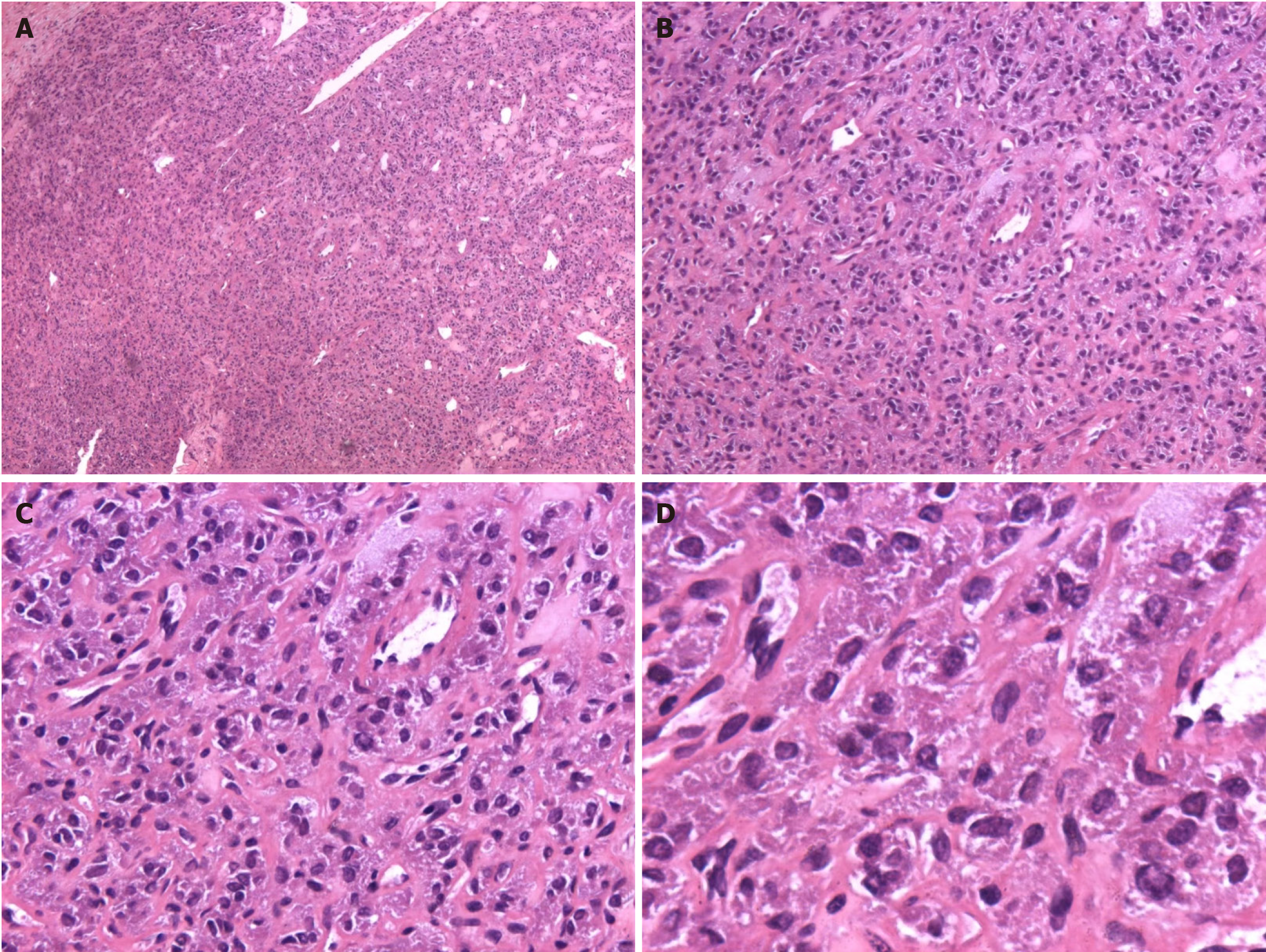
Figure 2 Postoperative microscopic images.
A: Heterogeneous cells arranged in organoid or chrysanthemum-shaped clusters [Hematoxylin and eosin (HE): 50 ×]; B: Cells with increased nucleoplasmic ratios and eosinophilic cytoplasm (HE: 100 ×); C: Interstitial fibrous tissue proliferation and myxoid degeneration (HE: 200 ×); D: Irregular nuclear membranes. finechromatin.no necrosis formation (HE: 400 ×).
- Citation: Luo SZ, Liu JR, Liu TQ, Chen Q. Functional paraganglioma of the pancreatic head: A case report and review of literature. World J Gastrointest Surg 2025; 17(7): 105833
- URL: https://www.wjgnet.com/1948-9366/full/v17/i7/105833.htm
- DOI: https://dx.doi.org/10.4240/wjgs.v17.i7.105833


