Copyright
©The Author(s) 2025.
World J Gastrointest Surg. Jul 27, 2025; 17(7): 104985
Published online Jul 27, 2025. doi: 10.4240/wjgs.v17.i7.104985
Published online Jul 27, 2025. doi: 10.4240/wjgs.v17.i7.104985
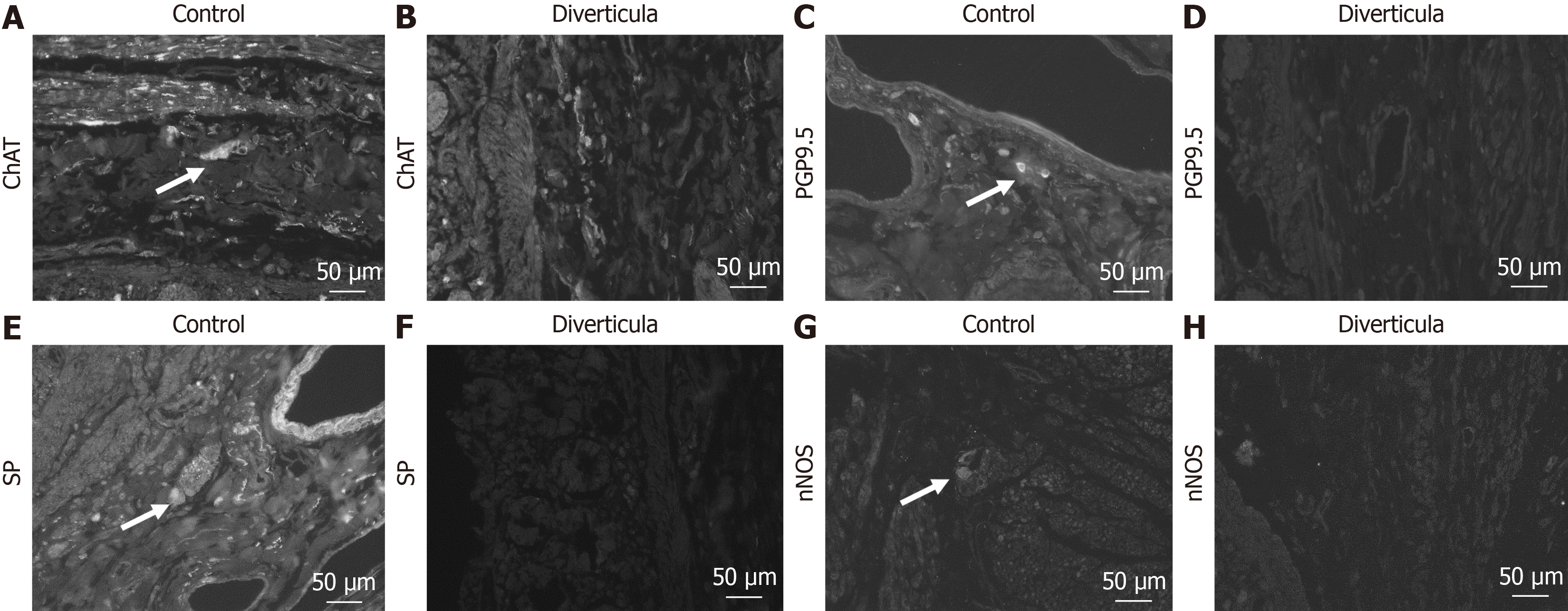
Figure 9 Immunohistochemical investigation of submucosal plexus in diverticula.
A-H: Detection of submucosal plexus in control tissue (A, C, E, and G) and diverticula (B, D, F, and H) utilizing choline acetyltransferase (ChAT) (A and B), protein gene product 9.5 (PGP9.5) (C and D), substance P (SP) (E and F) and neuronal nitric oxide synthase (nNOS) (G and H) antibody. Arrows mark positive cells in the plexus.
- Citation: Schmidt P, Perniss A, Nassenstein C, Keller H, Deckmann K. Multiple jejunal diverticulosis, from an anatomical and histological view: A case report. World J Gastrointest Surg 2025; 17(7): 104985
- URL: https://www.wjgnet.com/1948-9366/full/v17/i7/104985.htm
- DOI: https://dx.doi.org/10.4240/wjgs.v17.i7.104985


