Copyright
©The Author(s) 2025.
World J Gastrointest Surg. Jul 27, 2025; 17(7): 104985
Published online Jul 27, 2025. doi: 10.4240/wjgs.v17.i7.104985
Published online Jul 27, 2025. doi: 10.4240/wjgs.v17.i7.104985
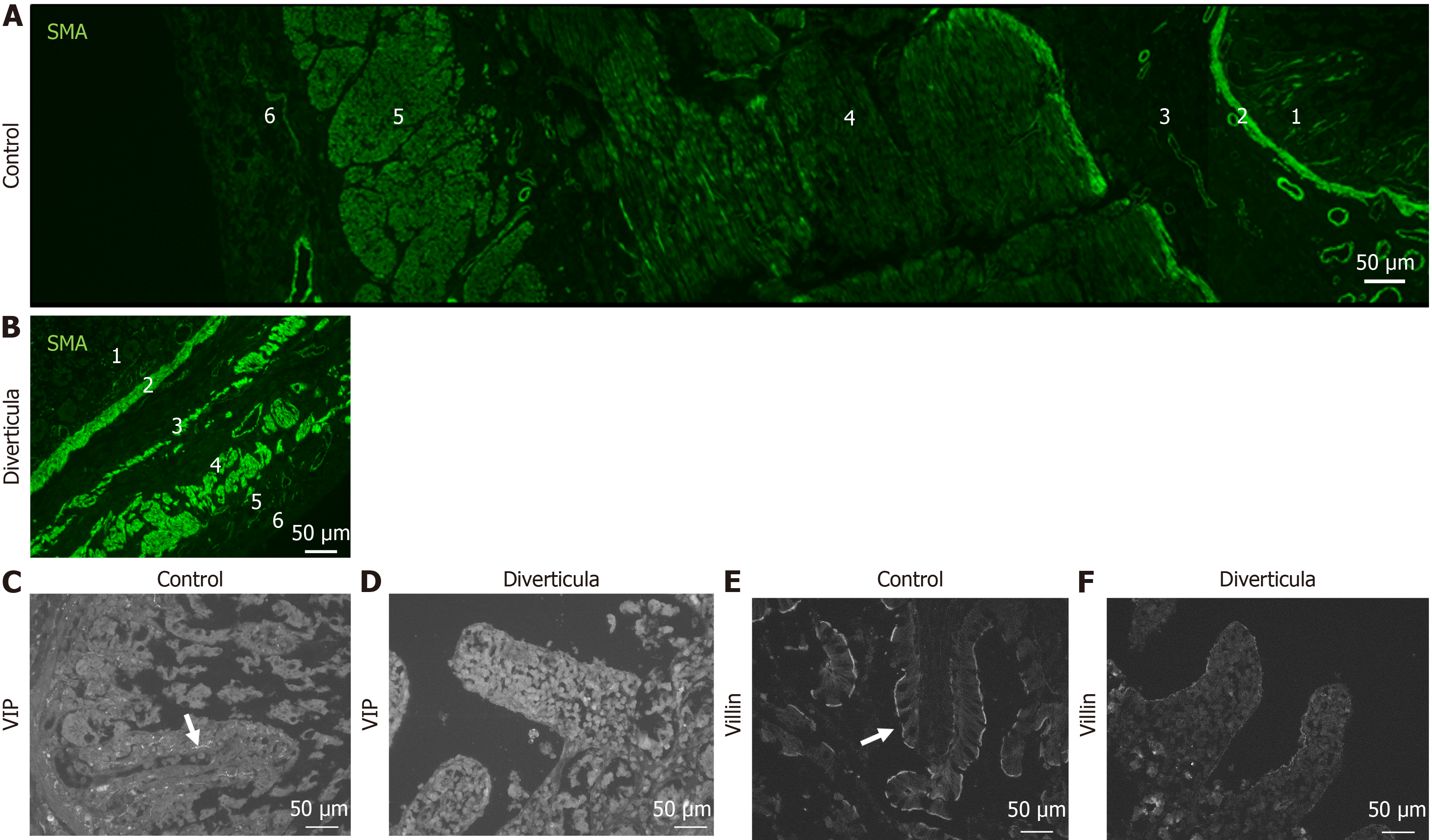
Figure 8 Immunohistochemical investigation of diverticula.
A and B: Immunohistochemical staining with smooth muscle actin antibody of control tissue (A) and diverticula (B); C-F: Investigation of intestinal villi in control tissue (C and E) and diverticula (D and F) utilizing vasoactive intestinal peptide (VIP; C and D) and villin (E and F) to mark VIP-positive fibers and brush border, respectively. 1: Intestinal villi; 2: Lamina muscularis mucosa; 3: Lamina submucosae; 4: Stratum circular; 5: Stratum longitudinale; 6: Adventitia/serosa.
- Citation: Schmidt P, Perniss A, Nassenstein C, Keller H, Deckmann K. Multiple jejunal diverticulosis, from an anatomical and histological view: A case report. World J Gastrointest Surg 2025; 17(7): 104985
- URL: https://www.wjgnet.com/1948-9366/full/v17/i7/104985.htm
- DOI: https://dx.doi.org/10.4240/wjgs.v17.i7.104985


