Copyright
©The Author(s) 2025.
World J Gastrointest Surg. Jul 27, 2025; 17(7): 104985
Published online Jul 27, 2025. doi: 10.4240/wjgs.v17.i7.104985
Published online Jul 27, 2025. doi: 10.4240/wjgs.v17.i7.104985
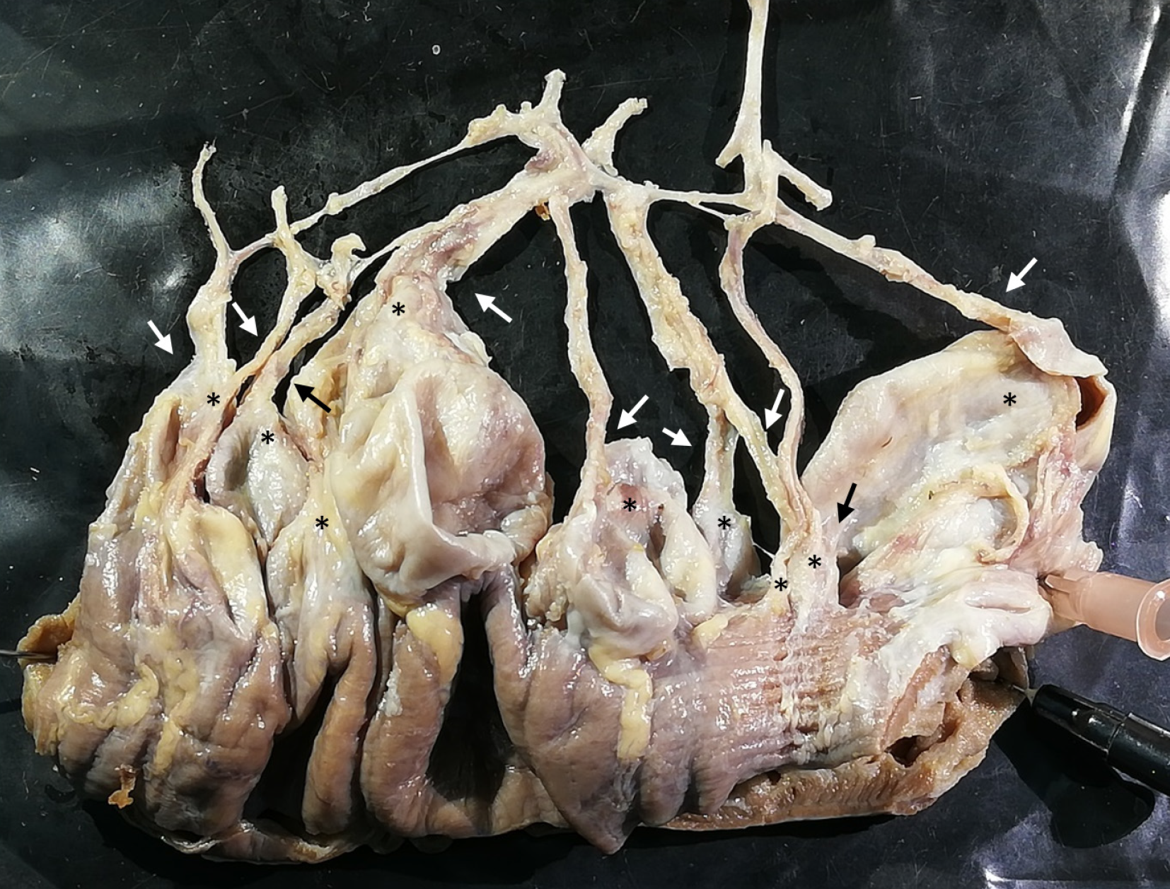
Figure 6 Relationship between vessels and diverticula.
Representative part of the jejunum (8 cm) with diverticula was cut out and dissected utilizing a dissecting microscope. All remaining fat tissue and intestine covering tunica serosa was removed, vessels were stretched out, and entry sites were exposed. Arrows mark arteria recta entering diverticula (marked with an asterisk). All investigated vessels end in diverticula, and all investigated diverticula were supplied by a vessel.
- Citation: Schmidt P, Perniss A, Nassenstein C, Keller H, Deckmann K. Multiple jejunal diverticulosis, from an anatomical and histological view: A case report. World J Gastrointest Surg 2025; 17(7): 104985
- URL: https://www.wjgnet.com/1948-9366/full/v17/i7/104985.htm
- DOI: https://dx.doi.org/10.4240/wjgs.v17.i7.104985


