Copyright
©The Author(s) 2025.
World J Gastrointest Surg. Jul 27, 2025; 17(7): 104777
Published online Jul 27, 2025. doi: 10.4240/wjgs.v17.i7.104777
Published online Jul 27, 2025. doi: 10.4240/wjgs.v17.i7.104777
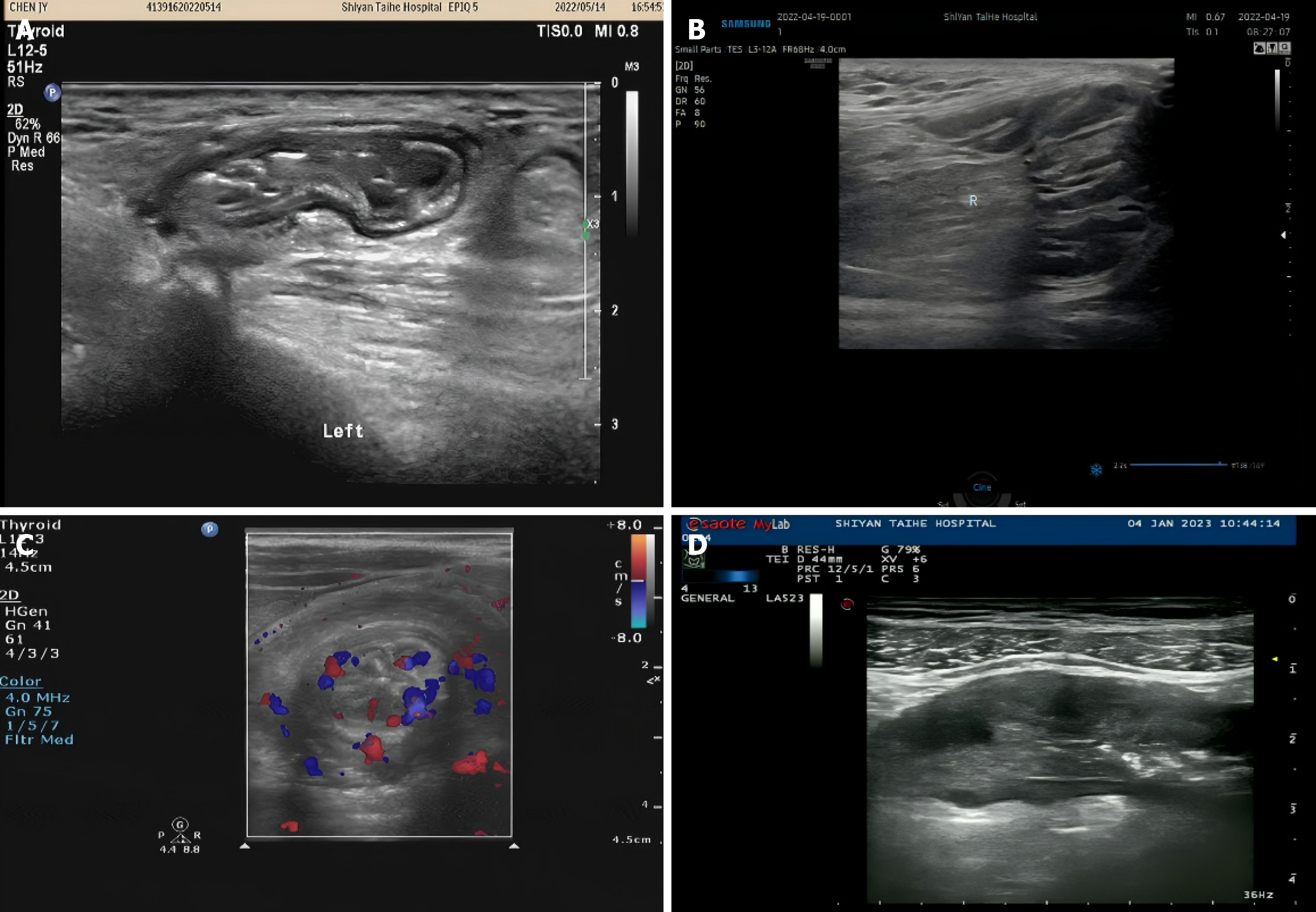
Figure 3 Severe adhesive ileus with complications.
A: Severe viscous ileus with internal hernia. The hernia ring structure is the adhered bowel, and the internal hernia shows the hernia ring structure and the bowel herniated into it; B: Severe adhesive ileus with intestinal volvulus. The root of the expanded loop is rotated; C: Severe adhesive ileus with intussusception. The short axis of the intestine is “concentric circle” sign, and the intestinal wall within the intussusception mass shows increased blood flow signal; D: Severe adhesive ileus with intestinal ischemic necrosis. The intestinal wall ischemia ultrasound shows that the layers of the ischemic intestinal wall are not clear.
- Citation: Wang F, Liu C, Wang H. Value analysis of ultrasound classification in disease judgment and treatment plan formulation of patients with adhesive intestinal obstruction. World J Gastrointest Surg 2025; 17(7): 104777
- URL: https://www.wjgnet.com/1948-9366/full/v17/i7/104777.htm
- DOI: https://dx.doi.org/10.4240/wjgs.v17.i7.104777


