Copyright
©The Author(s) 2025.
World J Gastrointest Surg. Jun 27, 2025; 17(6): 105007
Published online Jun 27, 2025. doi: 10.4240/wjgs.v17.i6.105007
Published online Jun 27, 2025. doi: 10.4240/wjgs.v17.i6.105007
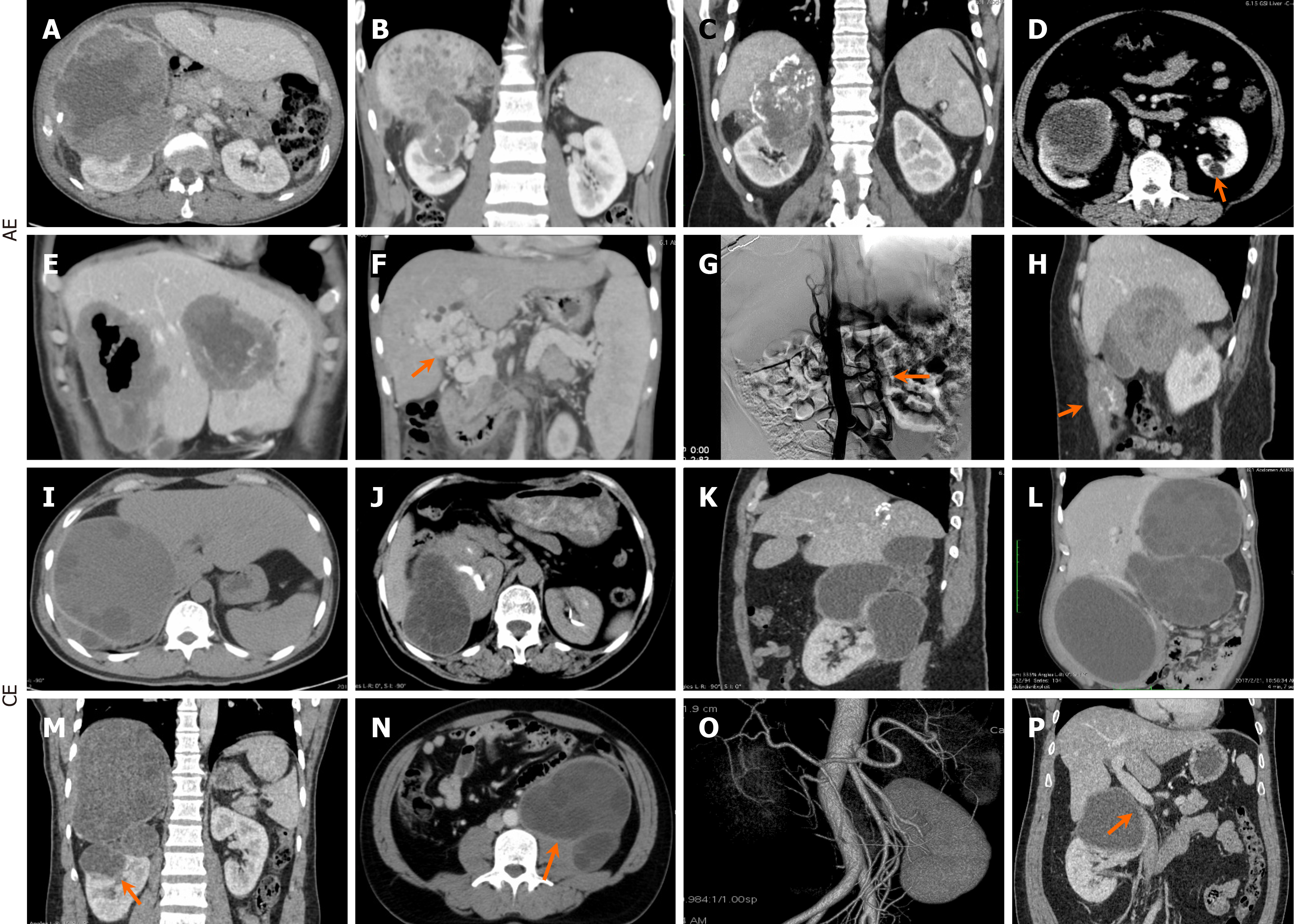
Figure 2 Radiological findings.
A-C: Right-sided hepatorenal alveolar echinococcosis (AE) lesion involvement was prevalent in AE cases; D and E: Isolated single lesions were observed in the left kidney (D) and the left lateral lobe of the liver (E); F-H: AE comorbidities included portal vein cavernous transformation (F), bilateral collateral circulation (G) secondary to severe inferior vena cava (IVC) involvement, and abdominal wall invasion (H); I-M: In contrast, cystic echinococcosis (CE) presented with solitary and multiple large cysts bilaterally; N-P: CE comorbidities included psoas muscle invasion (N), renal function impairment (O), and IVC involvement (P). AE: Alveolar echinococcosis; CE: Cystic echinococcosis.
- Citation: Tulahong A, Zhu DL, Liu C, Jiang TM, Zhang RQ, Tuergan T, Aji T, Shao YM. Simultaneous combined surgery for hepatic-renal double organ alveolar or cystic echinococcosis: A retrospective study. World J Gastrointest Surg 2025; 17(6): 105007
- URL: https://www.wjgnet.com/1948-9366/full/v17/i6/105007.htm
- DOI: https://dx.doi.org/10.4240/wjgs.v17.i6.105007


