Copyright
©The Author(s) 2025.
World J Gastrointest Surg. May 27, 2025; 17(5): 104893
Published online May 27, 2025. doi: 10.4240/wjgs.v17.i5.104893
Published online May 27, 2025. doi: 10.4240/wjgs.v17.i5.104893
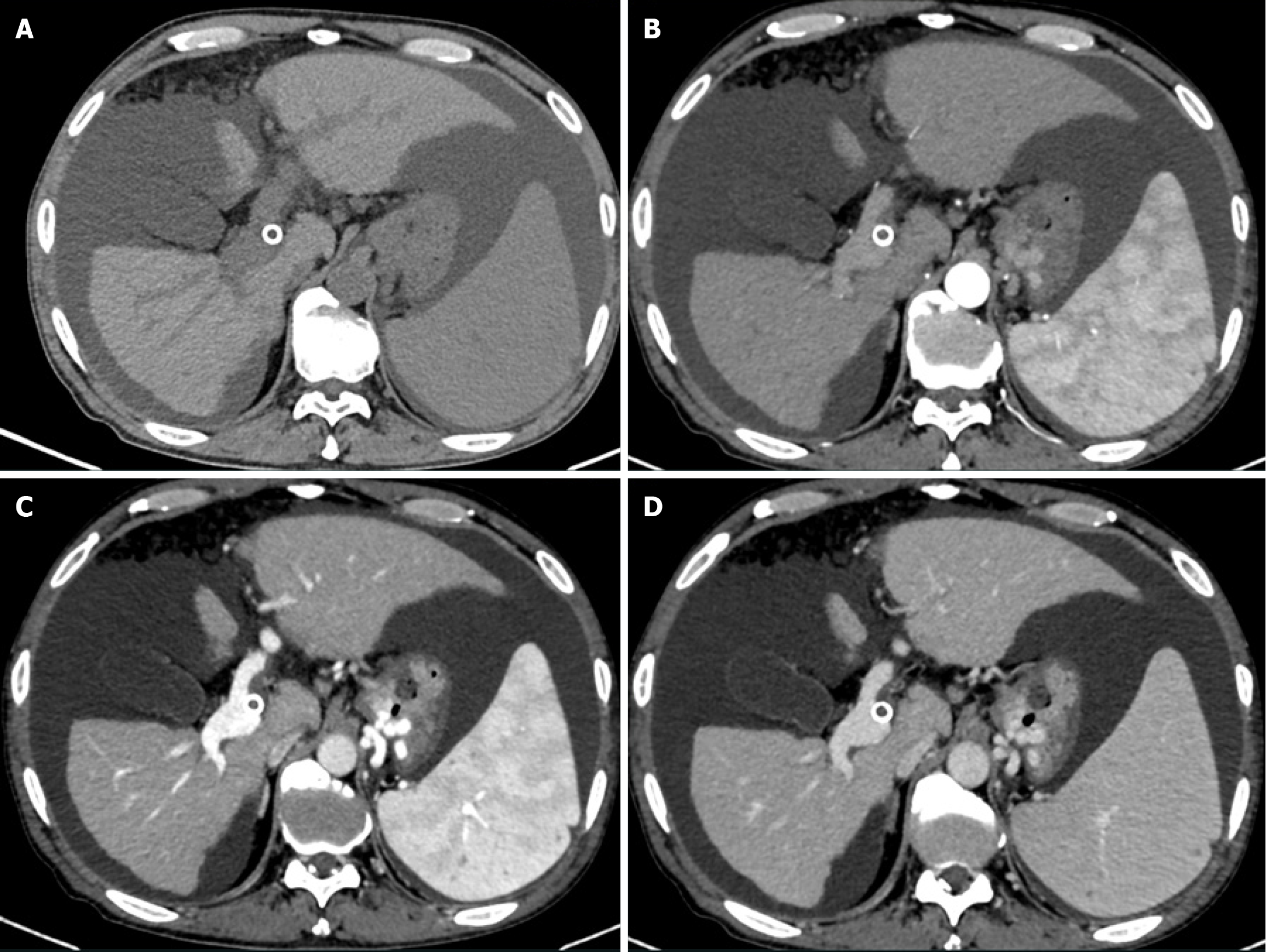
Figure 4 Thirty-month follow-up computed tomography after transjugular intrahepatic portosystemic shunt.
Computed tomography demonstrates shunt thrombosis accompanied by recurrent esophagogastric varices and massive ascites. A: Unenhanced phase; B: Hepatic arterial phase; C: Portal venous phase; D: Delayed phase.
- Citation: Zhou TY, Wang HL, Tao GF, Chen SQ. Stent fracture after transjugular intrahepatic portosystemic shunt: A case report. World J Gastrointest Surg 2025; 17(5): 104893
- URL: https://www.wjgnet.com/1948-9366/full/v17/i5/104893.htm
- DOI: https://dx.doi.org/10.4240/wjgs.v17.i5.104893


