
















©The Author(s) 2025.
World J Gastrointest Surg. Mar 27, 2025; 17(3): 102174
Published online Mar 27, 2025. doi: 10.4240/wjgs.v17.i3.102174
Published online Mar 27, 2025. doi: 10.4240/wjgs.v17.i3.102174
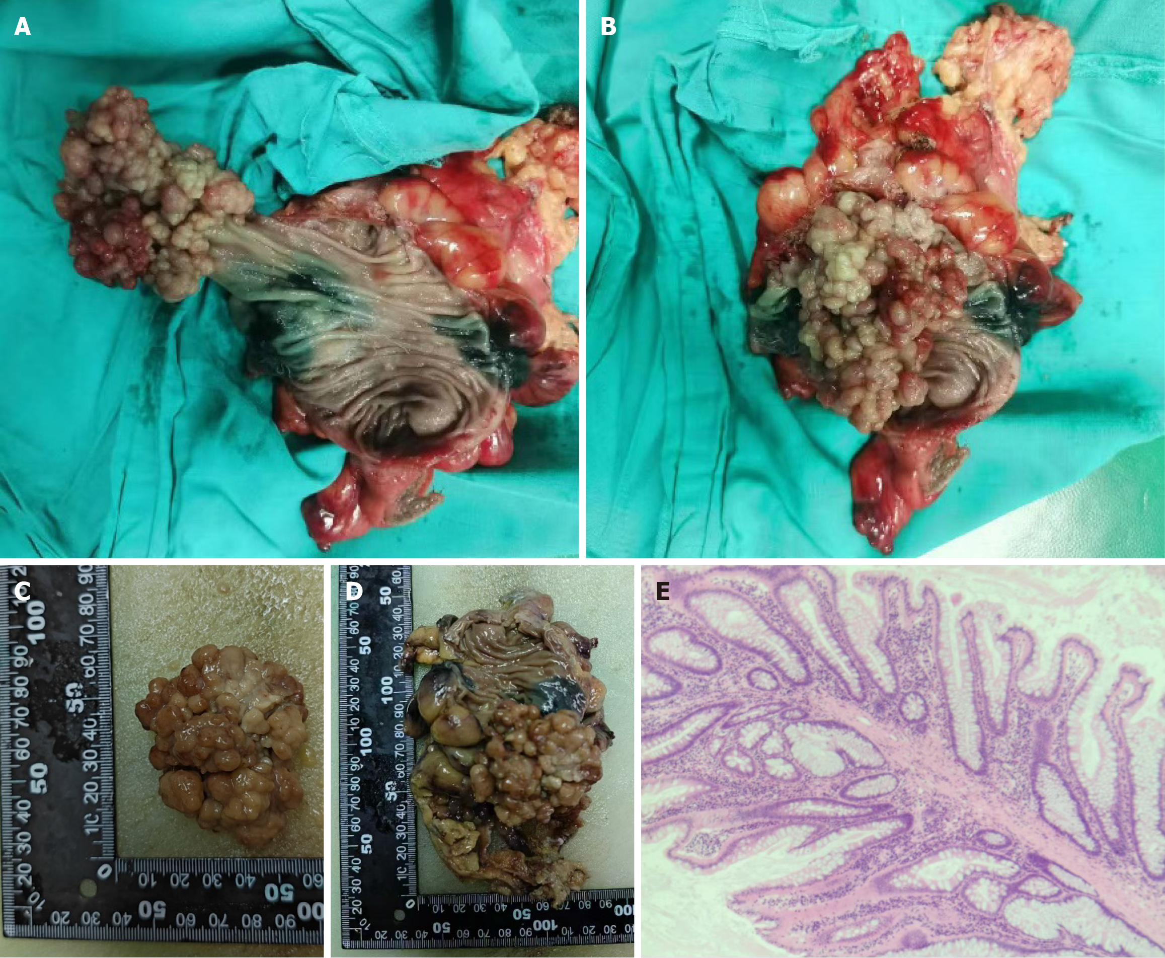
Figure 5 Postoperative tissue sample and pathological results.
A-D: The resected grape-like mass of sigmoid colon was showed and the polyp size was approximately 6 cm × 5 cm × 5 cm; E: Hematoxylin-eosin staining of the lesions. There were dendritic-like structures formed by the proliferation of muscle fibers in the muscularis mucosa, which were overlaid with intrinsic mucosa tissue and piled up into villous structures.
- Citation: Tian ZS, Ma XP, Ruan HX, Yang Y, Zhao YL. Rare large sigmoid hamartomatous polyp in an elderly patient with atypical Peutz-Jeghers syndrome: A case report. World J Gastrointest Surg 2025; 17(3): 102174
- URL: https://www.wjgnet.com/1948-9366/full/v17/i3/102174.htm
- DOI: https://dx.doi.org/10.4240/wjgs.v17.i3.102174