
















©The Author(s) 2025.
World J Gastrointest Surg. Dec 27, 2025; 17(12): 113532
Published online Dec 27, 2025. doi: 10.4240/wjgs.v17.i12.113532
Published online Dec 27, 2025. doi: 10.4240/wjgs.v17.i12.113532
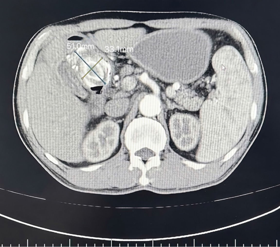
Figure 1 This computed tomography image is the abdominal cross-sectional image of this patient on March 22, 2022.
In the image, an oval high-density gallstone (about 5.0 cm in diameter) was embedded in the proximal duodenum, resulting in significant expansion of the stomach. Edema of duodenal wall and local intestinal lumen stenosis can be seen around the stone. In addition, the gas shadow in the gallbladder suggests that there may be abnormal channels between the gallbladder floor and the gastric antrum, with a little exudation around the gastric antrum.
- Citation: Hu YC, Chen XY, Cao MK, Fan Z. Bouveret syndrome in a young patient: A case report and review of literature. World J Gastrointest Surg 2025; 17(12): 113532
- URL: https://www.wjgnet.com/1948-9366/full/v17/i12/113532.htm
- DOI: https://dx.doi.org/10.4240/wjgs.v17.i12.113532