
















©The Author(s) 2025.
World J Gastrointest Surg. Dec 27, 2025; 17(12): 112887
Published online Dec 27, 2025. doi: 10.4240/wjgs.v17.i12.112887
Published online Dec 27, 2025. doi: 10.4240/wjgs.v17.i12.112887
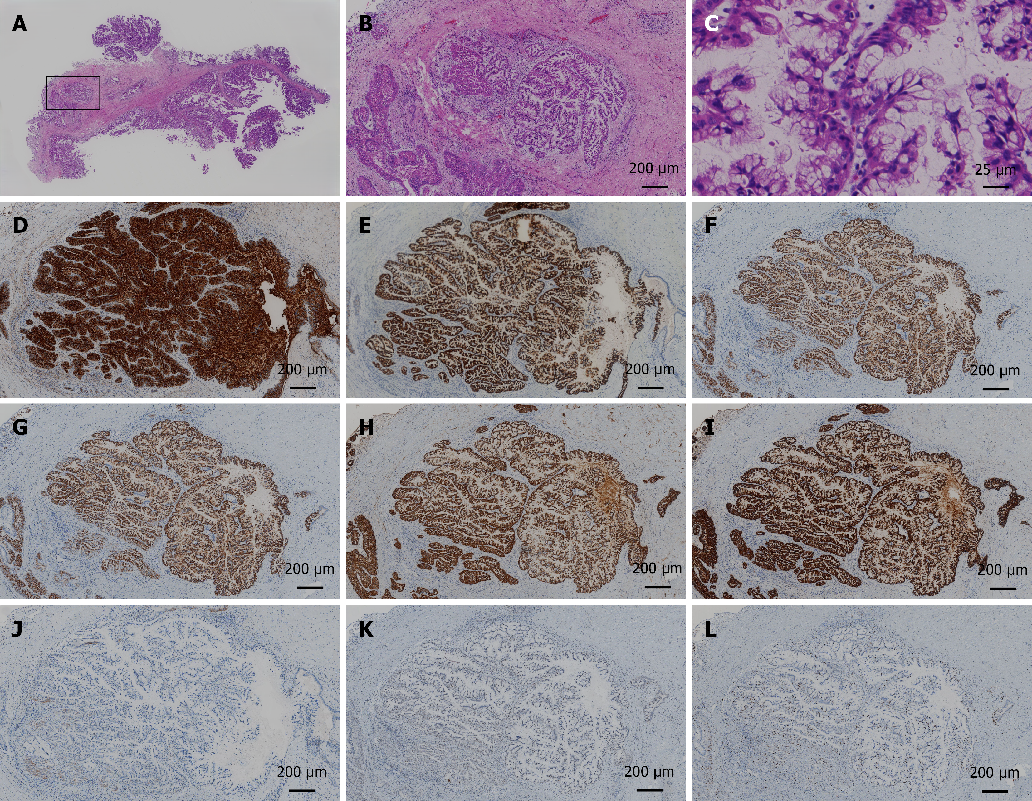
Figure 4 Histopathologic features of the surgical specimen.
A-C: Haematoxylin and eosin staining of the papillary tumour: Macrography (A); × 40 (B); × 400 (C); D-K: Immunohistochemical findings of the papillary tumour: MUC1 positivity (× 40; D); MUC2 positivity (× 40; E); MUC5AC positivity (× 40; F); MUC6 positivity (× 40; G); CK7 positivity (× 40; H); CK19 positivity (× 40; I); CK20 negativity (× 40; J); P53 wild-type (× 40; K); L: Ki-67 expression in approximately 20% of the cells, indicating moderate proliferative activity (× 40).
- Citation: Gu TY, Wang SH, Yang SY, Zhao SQ, Chen MJ, Li XP, Song ZW, Gu XX, Chen F. Intraductal papillary neoplasm of the biliary tract with typical clinicopathological, endoscopic features: A case report. World J Gastrointest Surg 2025; 17(12): 112887
- URL: https://www.wjgnet.com/1948-9366/full/v17/i12/112887.htm
- DOI: https://dx.doi.org/10.4240/wjgs.v17.i12.112887