
















©The Author(s) 2025.
World J Gastrointest Surg. Dec 27, 2025; 17(12): 112887
Published online Dec 27, 2025. doi: 10.4240/wjgs.v17.i12.112887
Published online Dec 27, 2025. doi: 10.4240/wjgs.v17.i12.112887
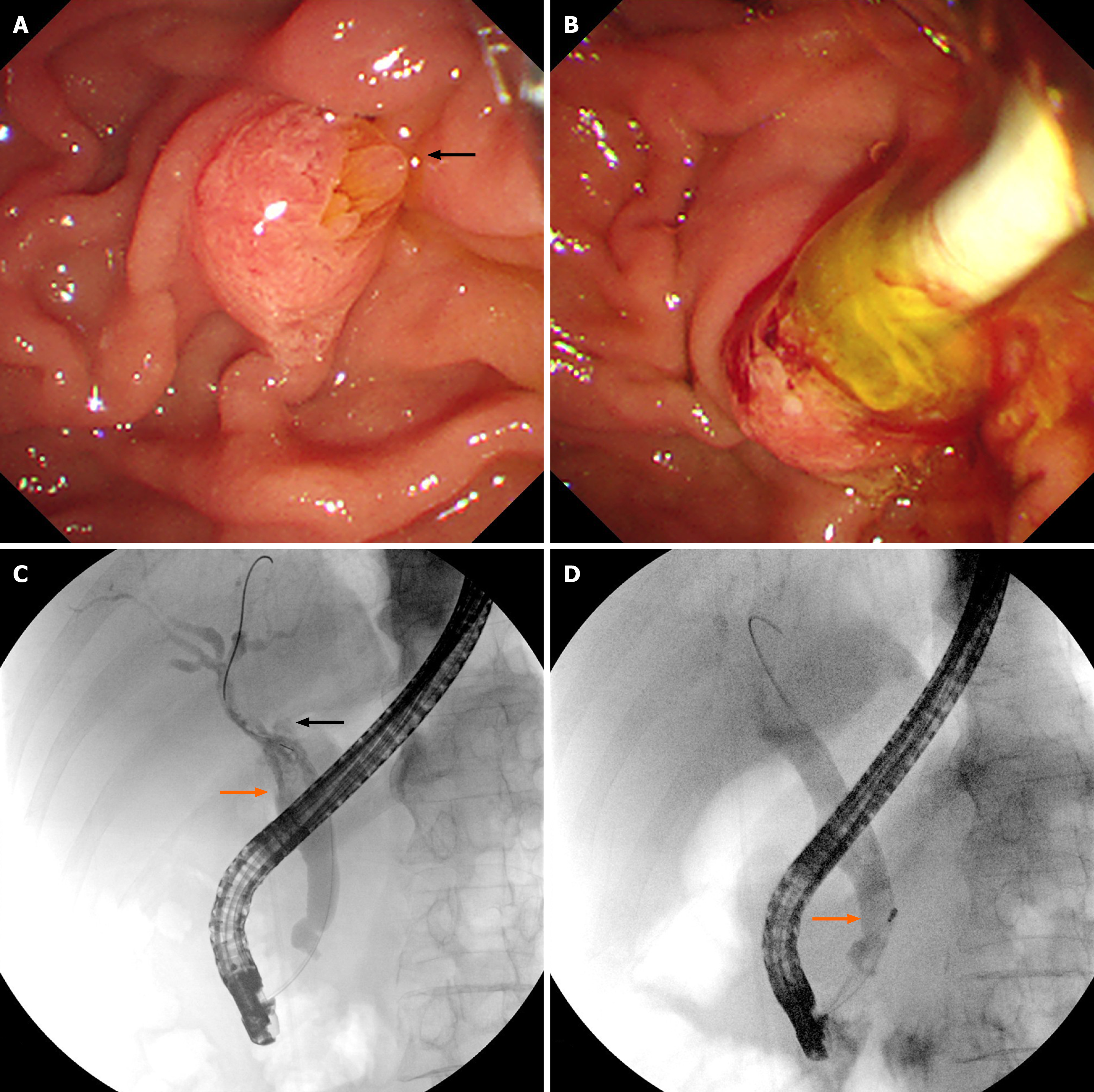
Figure 2 Endoscopic retrograde cholangiopancreatography findings.
A: Duodenoscopy revealed an enlarged duodenal papilla, a dilated papillary orifice with a fish-mouth-like appearance and translucent jelly mucus (black arrow); B: Duodenoscopy revealed a large amount of translucent jelly-like mucus containing bile flowing out during bile duct clearance; C: Endoscopic retrograde cholangiography showed multiple cloudy, irregular filling defects in the dilated extrahepatic bile duct (orange arrow), non-visualized dilated left hepatic duct and an intrahepatic cystic lesion caused by mucin obstruction (black arrow); D: Endoscopic retrograde cholangiography showed the morphology and location of the filling defect in the bile duct were altered during clearing of the bile duct (orange arrow).
- Citation: Gu TY, Wang SH, Yang SY, Zhao SQ, Chen MJ, Li XP, Song ZW, Gu XX, Chen F. Intraductal papillary neoplasm of the biliary tract with typical clinicopathological, endoscopic features: A case report. World J Gastrointest Surg 2025; 17(12): 112887
- URL: https://www.wjgnet.com/1948-9366/full/v17/i12/112887.htm
- DOI: https://dx.doi.org/10.4240/wjgs.v17.i12.112887