
















©The Author(s) 2025.
World J Gastrointest Surg. Dec 27, 2025; 17(12): 110617
Published online Dec 27, 2025. doi: 10.4240/wjgs.v17.i12.110617
Published online Dec 27, 2025. doi: 10.4240/wjgs.v17.i12.110617
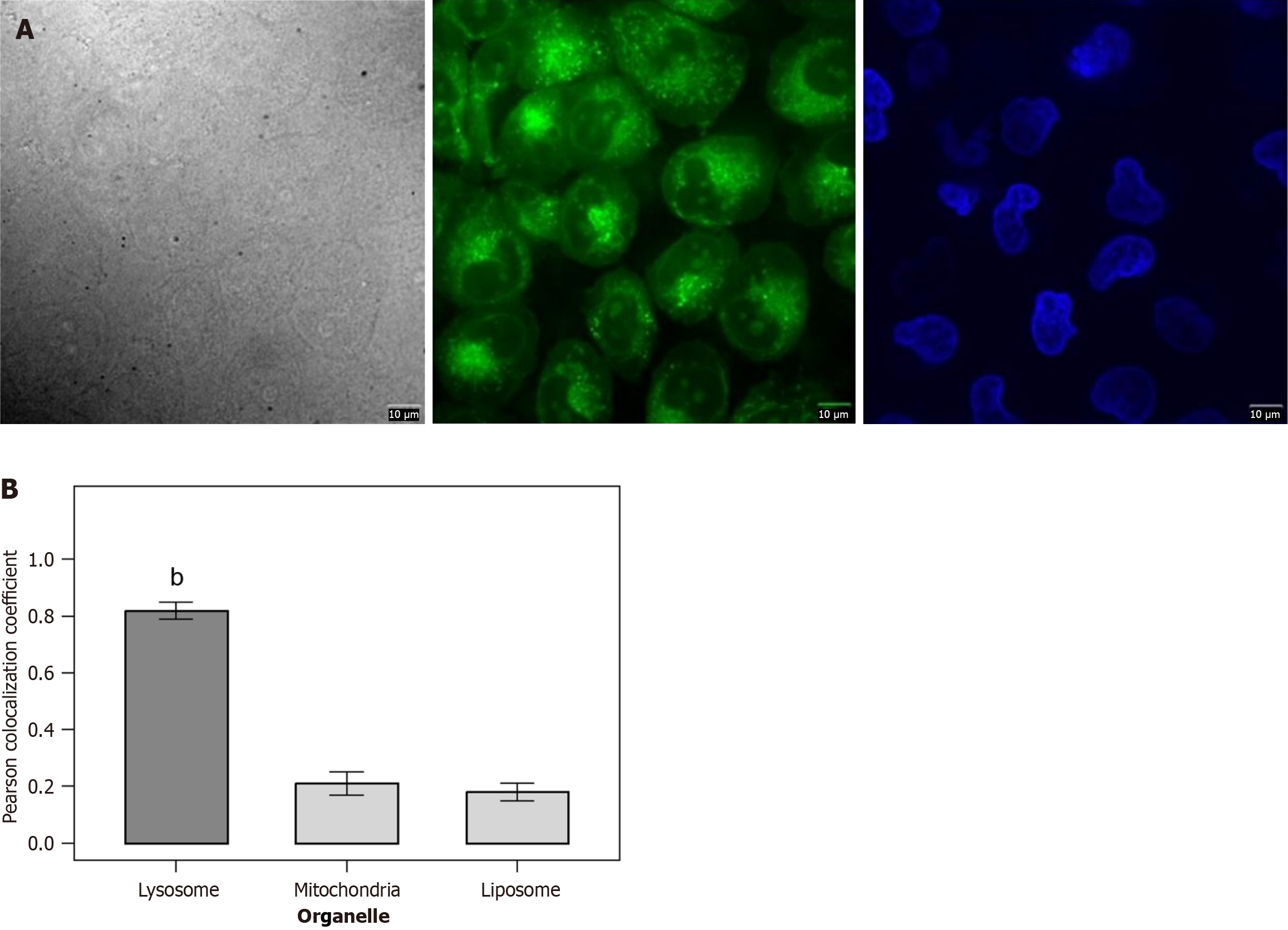
Figure 4 Targeted colocalization of DAF-FM probe with lysosomes.
A: Merged images of DAF-FM (red) with Lyso-Tracker Green (lysosome, green), Mito-Tracker Green (mitochondria, green), and BODIPY493/593 (liposome, green); B: Bar chart of Pearson colocalization coefficients (lysosome: 0.82 ± 0.03, mitochondria: 0.21 ± 0.04, liposome: 0.18 ± 0.03). Scale bar: 10 μm; data are expressed as mean ± SD, n = 3 per group, bP < 0.01 vs mitochondria/Liposome groups.
- Citation: Chen WH, Cai CF, Gao BZ, Hong WS, Xu YZ, Cai WJ. Visual screening and efficacy evaluation of high-performance fluorescent probe DAF-FM in esophagitis cancer transformation. World J Gastrointest Surg 2025; 17(12): 110617
- URL: https://www.wjgnet.com/1948-9366/full/v17/i12/110617.htm
- DOI: https://dx.doi.org/10.4240/wjgs.v17.i12.110617