
















©The Author(s) 2025.
World J Gastrointest Surg. Dec 27, 2025; 17(12): 110617
Published online Dec 27, 2025. doi: 10.4240/wjgs.v17.i12.110617
Published online Dec 27, 2025. doi: 10.4240/wjgs.v17.i12.110617
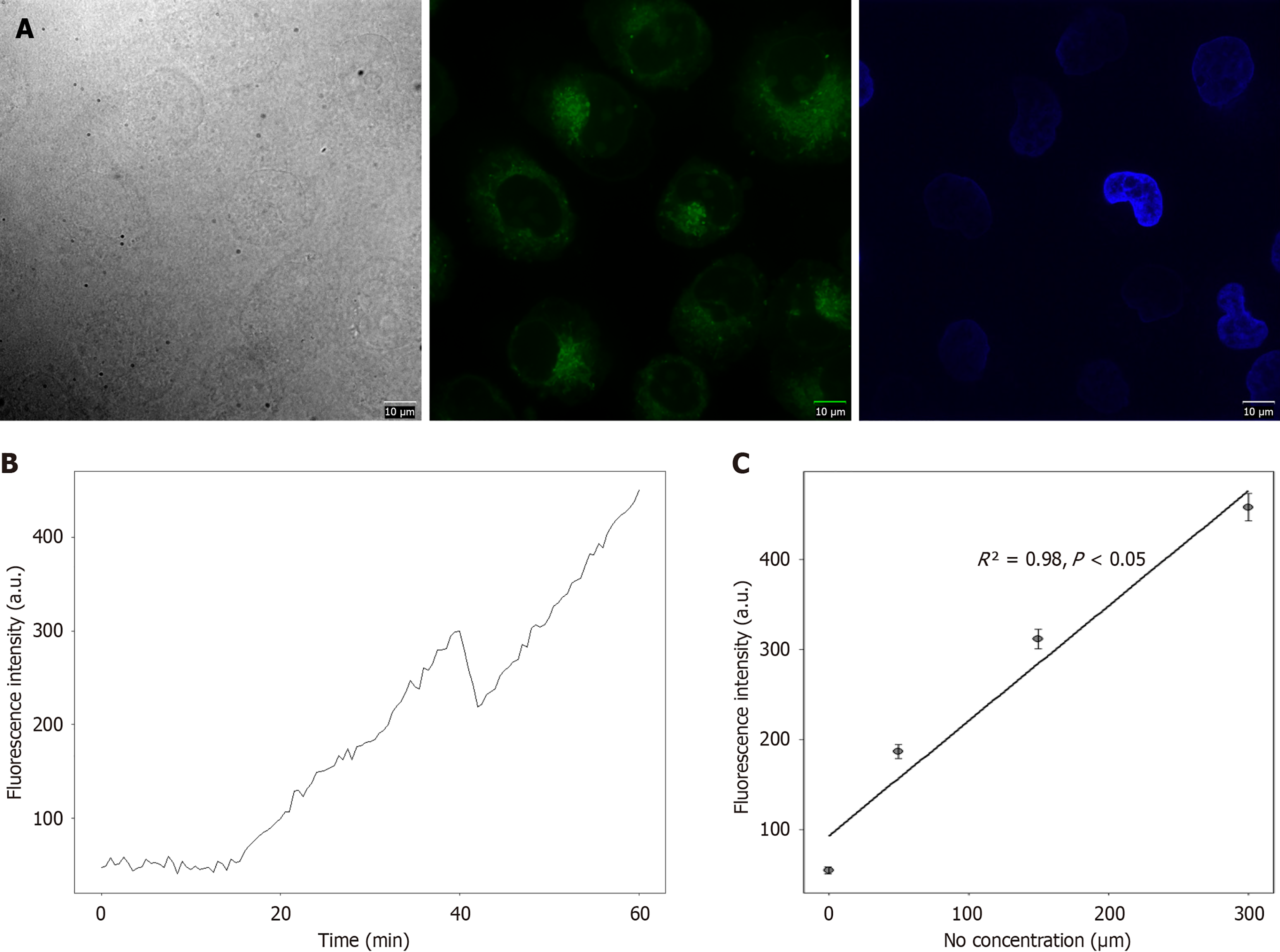
Figure 2 Concentration-dependent and time-dependent fluorescence intensity of DAF-FM probe with nitric oxide.
A: Fluorescence images of DAF-FM incubated with different concentrations of nitric oxide (NO) donor DEANO (0 μM, 50 μM, 150 μM, 300 μM) for 60 minutes; B: Fluorescence intensity-time curve (0–60 minutes) of DAF-FM at different NO concentrations [0 μM: From 52 ± 3 to 55 ± 4 arbitrary units (a.u.); 300 μM: From 223 ± 9 to 458 ± 15 a.u.]; C: Fluorescence intensity-concentration scatter plot of DAF-FM at 60 minutes (R² = 0.98, P < 0.05). Scale bar: 10 μm; data are expressed as mean ± SD, n = 3 per group.
- Citation: Chen WH, Cai CF, Gao BZ, Hong WS, Xu YZ, Cai WJ. Visual screening and efficacy evaluation of high-performance fluorescent probe DAF-FM in esophagitis cancer transformation. World J Gastrointest Surg 2025; 17(12): 110617
- URL: https://www.wjgnet.com/1948-9366/full/v17/i12/110617.htm
- DOI: https://dx.doi.org/10.4240/wjgs.v17.i12.110617