
















©The Author(s) 2025.
World J Gastrointest Surg. Nov 27, 2025; 17(11): 110864
Published online Nov 27, 2025. doi: 10.4240/wjgs.v17.i11.110864
Published online Nov 27, 2025. doi: 10.4240/wjgs.v17.i11.110864
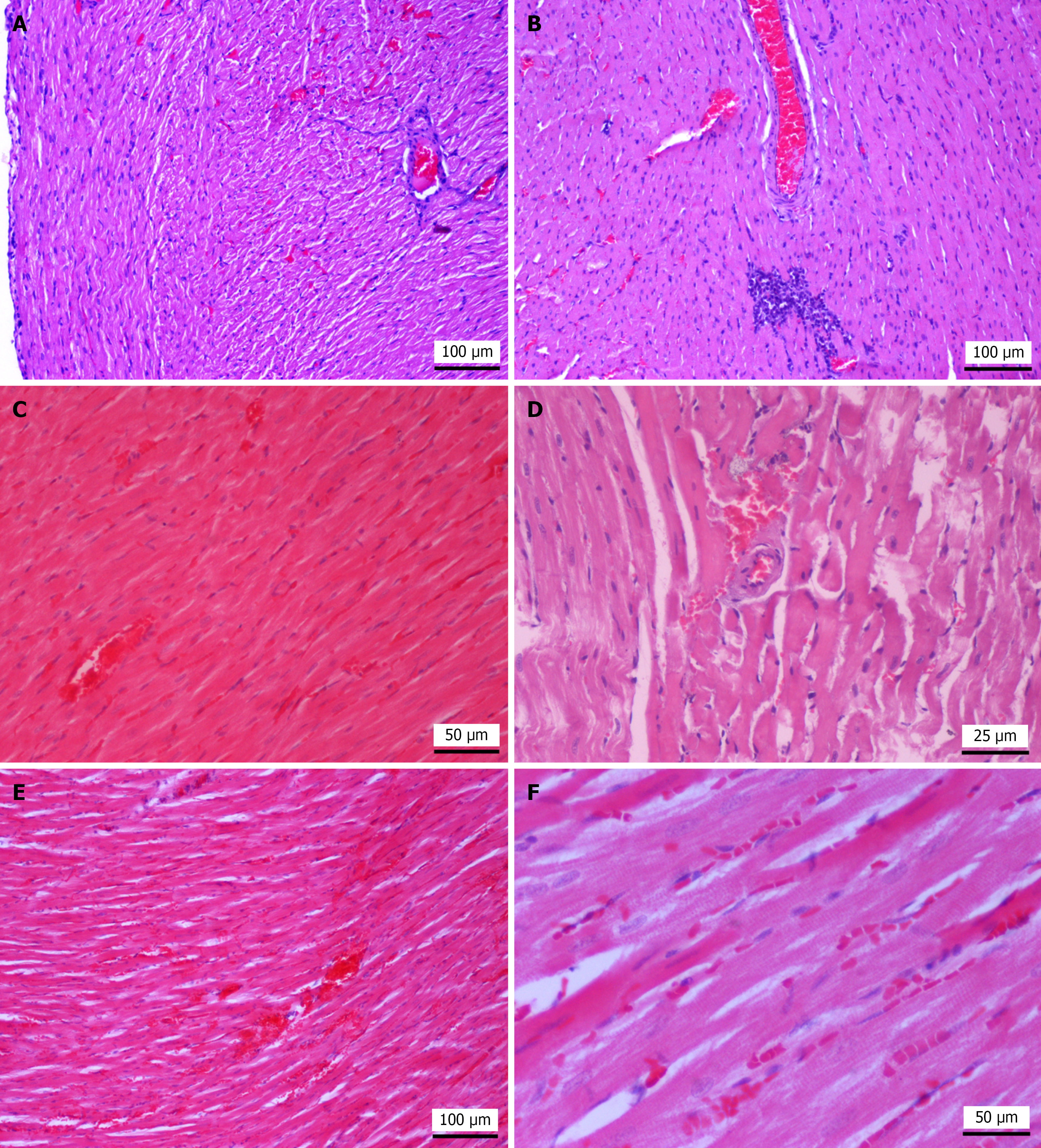
Figure 6 Representative histopathological images of heart tissue in sham group, hepatic ischemia-reperfusion injury group, treprostinil treated group, and ischemic preconditioning group.
A: Sham group; the myocardium in a rat from the sham group shows well-preserved cardiomyocytes. Mild interstitial edema, sinusoidal hyperhemia and presence of wavy fibers located subepicardially are also observed. Hematoxylin and eosin (HE), original magnification, 100 ×; B: Hepatic ischemia-reperfusion injury group (HIRI); rat subjected to HIRI shows loss of striation with homogenously eosinophilic sarcoplasm, a large mononuclear cell aggregate and pyknotic nuclei (necrosis). HE, original magnification, 100 ×; C: Treprostinil-treated group; treprostinil-treated rat shows mild edema and cardiomyocyte necrosis. HE, original magnification, 200 ×; D: Ischemic preconditioning (IP) group; rats subjected to hepatic IP show mild interstitial edema, segmental loss of striation and eosinophilic sarcoplasm. HE, original magnification, 400 ×; E: IP group, rats subjected to hepatic IP show hyperhemia, hemorrhage and edema. HE, original magnification, 100 ×; F: IP group; rats subjected to hepatic IP show eosinophilic sarcoplasm, pyknotic nuclei necrosis and presence of sparse mononuclear cells. HE, original magnification, 200 ×.
- Citation: Mouratidou C, Pavlidis ET, Katsanos G, Kotoulas SC, Papaioannou M, Tsoulfas G, Apostolopoulou E, Brellou GD, Galanis IN, Pavlidis TE. Protective effects of treprostinil and ischemic preconditioning on hepatic ischemia-reperfusion injury and biomarkers in experimental studies in rats. World J Gastrointest Surg 2025; 17(11): 110864
- URL: https://www.wjgnet.com/1948-9366/full/v17/i11/110864.htm
- DOI: https://dx.doi.org/10.4240/wjgs.v17.i11.110864