
















©The Author(s) 2025.
World J Gastrointest Surg. Nov 27, 2025; 17(11): 110075
Published online Nov 27, 2025. doi: 10.4240/wjgs.v17.i11.110075
Published online Nov 27, 2025. doi: 10.4240/wjgs.v17.i11.110075
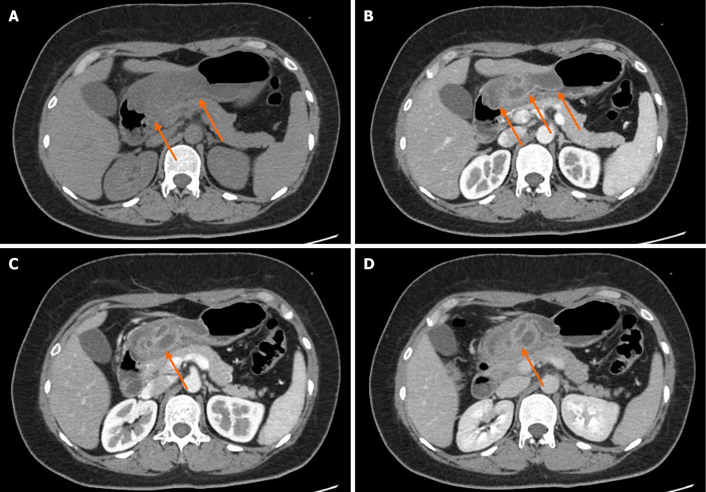
Figure 2 Contrast-enhanced computed tomography images.
A: Plain computed tomography showed significant thickening of the anterior wall of the gastric body and antrum with low-density areas; B: The mucosal line appeared continuous, with the lesion in the submucosa surrounded by patchy edema; C: In the arterial phase, multiple submucosal cystic lesions of varying sizes were visible in the anterior wall of the gastric antrum, with ring-like enhancement of the cyst walls; D: In the venous phase, the cyst walls exhibited persistent enhancement; in contrast, the cyst contents remained non-enhancing.
- Citation: Cui Q, He K, Chen MS, Huang LD. Diagnostic challenge of gastritis cystica profunda with secondary abscess formation: A case report. World J Gastrointest Surg 2025; 17(11): 110075
- URL: https://www.wjgnet.com/1948-9366/full/v17/i11/110075.htm
- DOI: https://dx.doi.org/10.4240/wjgs.v17.i11.110075