
















©The Author(s) 2025.
World J Gastrointest Surg. Nov 27, 2025; 17(11): 110075
Published online Nov 27, 2025. doi: 10.4240/wjgs.v17.i11.110075
Published online Nov 27, 2025. doi: 10.4240/wjgs.v17.i11.110075
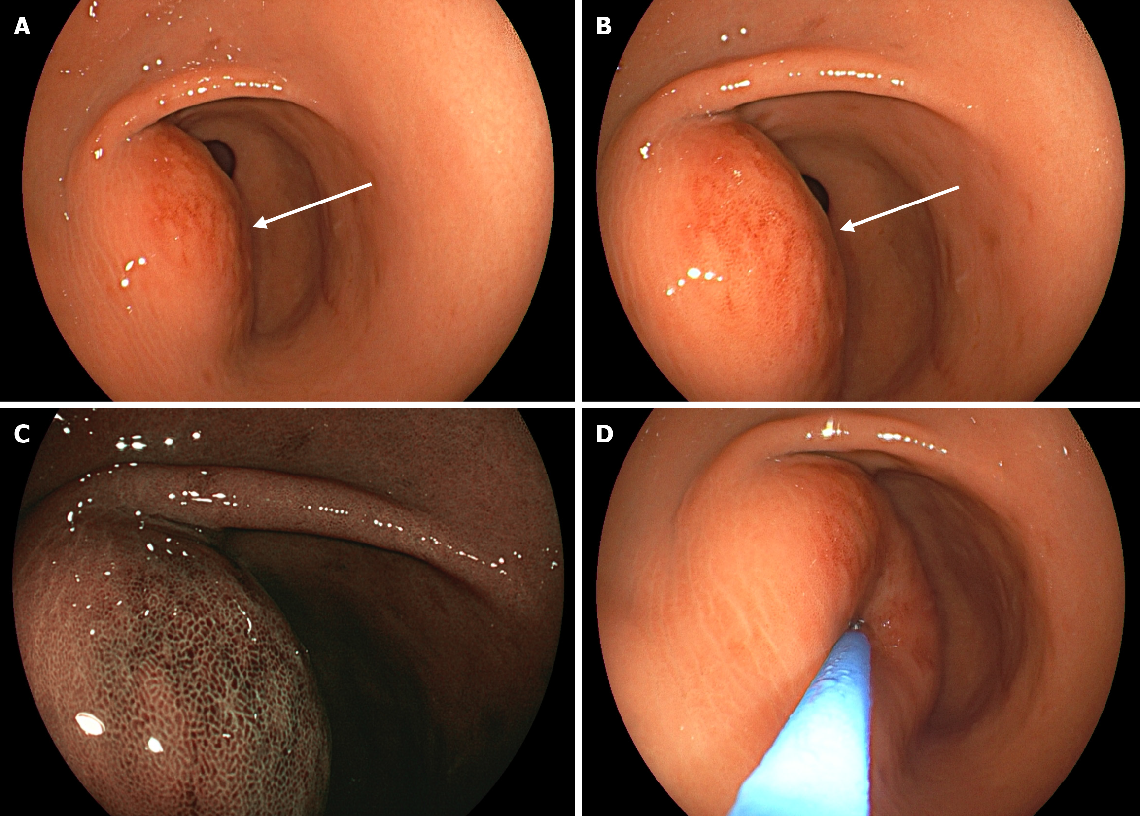
Figure 1 Gastroscopy showing a submucosal bulge in the gastric antrum.
A: White-light gastroscopy revealed congestion and edema of the gastric antral mucosa, with a 4-cm submucosal bulge on the anterior wall; B: Close-up view of the lesion; C: Optical electronic chromoendoscopy demonstrated the micro-surface and microvascular structures of the lesion; D: Application of pressure with a pair of biopsy forceps confirmed a soft texture.
- Citation: Cui Q, He K, Chen MS, Huang LD. Diagnostic challenge of gastritis cystica profunda with secondary abscess formation: A case report. World J Gastrointest Surg 2025; 17(11): 110075
- URL: https://www.wjgnet.com/1948-9366/full/v17/i11/110075.htm
- DOI: https://dx.doi.org/10.4240/wjgs.v17.i11.110075