
















©The Author(s) 2025.
World J Gastrointest Surg. Oct 27, 2025; 17(10): 111975
Published online Oct 27, 2025. doi: 10.4240/wjgs.v17.i10.111975
Published online Oct 27, 2025. doi: 10.4240/wjgs.v17.i10.111975
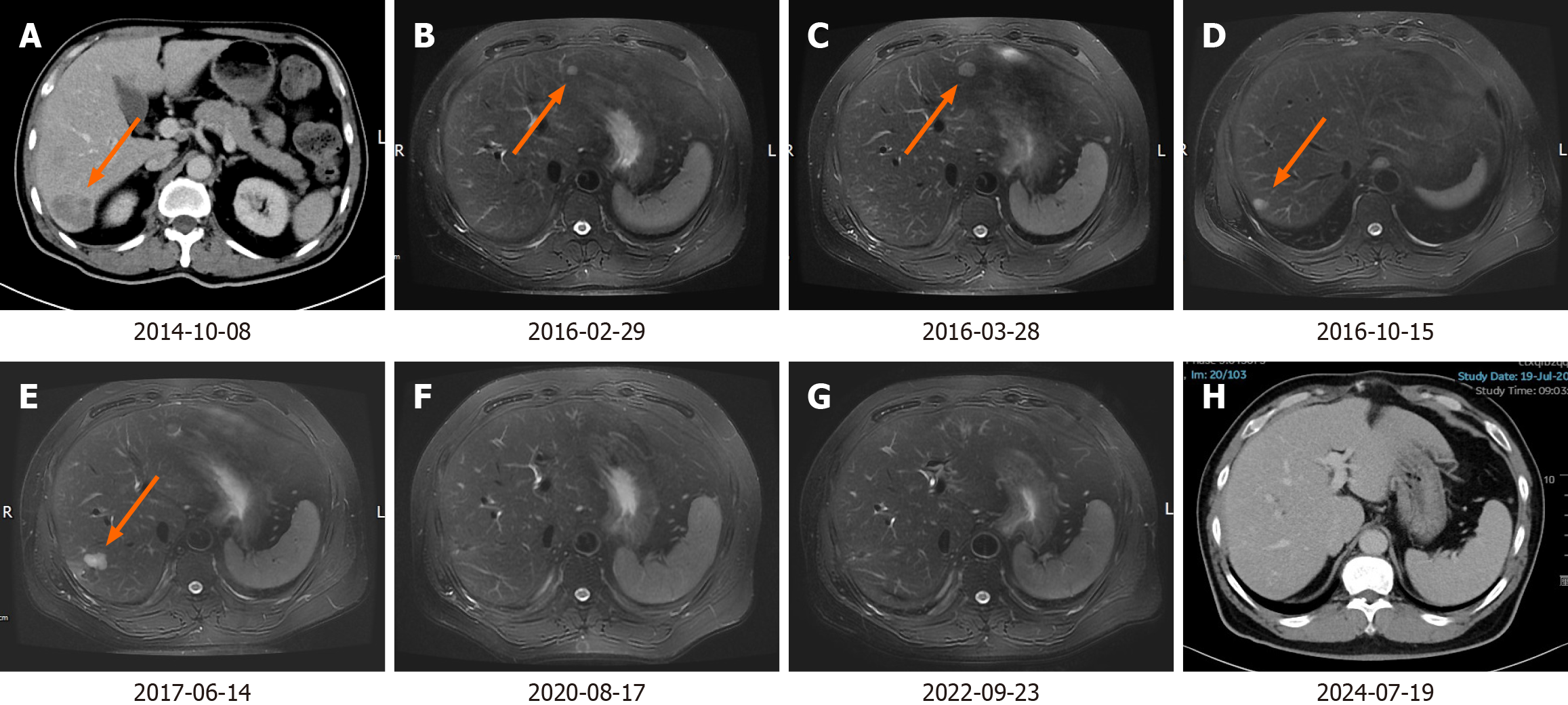
Figure 2 Abdominal computed tomography and magnetic resonance during the treatment and follow-up.
A: On October 8, 2014, a nodular lesion was found in the segment VI of the liver; B: On February 29, 2016, punctate and small nodular abnormal signals were found in both the lobes of the liver; C: On May 28, 2016, the size of the original punctate and small nodular abnormal signals in the liver had increased; D: On October 15, 2016, a new lesion in the segment VI of the liver was found; E: On June 14, 2017, several new nodules were found in the right lobe of the liver; F: On August 17, 2020, reexamination showed no new lesions in the liver; G: On September 23, 2022, reexamination showed no new lesions in the liver; H: On July 19, 2024, reexamination showed no new lesions in the liver. Orange arrows indicate the main lesions.
- Citation: Pan J, Zhang RS, Chen Y, Cao XH, Yang ZH, Lu L, Chen YT, Chu XY. Long-term survival after multimodality treatment of metastatic mixed adenoneuroendocrine carcinoma of colon: A case report. World J Gastrointest Surg 2025; 17(10): 111975
- URL: https://www.wjgnet.com/1948-9366/full/v17/i10/111975.htm
- DOI: https://dx.doi.org/10.4240/wjgs.v17.i10.111975