
















©The Author(s) 2025.
World J Gastrointest Surg. Oct 27, 2025; 17(10): 111975
Published online Oct 27, 2025. doi: 10.4240/wjgs.v17.i10.111975
Published online Oct 27, 2025. doi: 10.4240/wjgs.v17.i10.111975
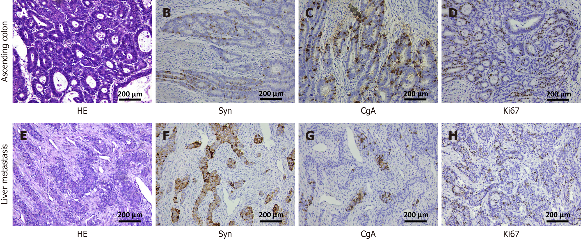
Figure 1 Hematoxylin and eosin and immunohistochemistry staining of ascending colon and liver metastasis.
A: Hematoxylin and eosin (HE) staining of mixed adenoneuroendocrine carcinoma (MANEC) of ascending colon; B: Immunohistochemistry (IHC) staining of ascending colon showed Syn (++); C: IHC staining of ascending colon showed CgA (+); D: IHC staining of ascending colon showed Ki67 (90%+); E: HE staining of MANEC of liver metastasis; F: IHC staining of liver metastasis showed Syn (++); G: IHC staining of liver metastasis showed CgA (+); H: IHC staining of liver metastasis showed Ki67 (90%+) ( × 100). HE: Hematoxylin and eosin; Syn: Synaptophysin; CgA: Chromogranin.
- Citation: Pan J, Zhang RS, Chen Y, Cao XH, Yang ZH, Lu L, Chen YT, Chu XY. Long-term survival after multimodality treatment of metastatic mixed adenoneuroendocrine carcinoma of colon: A case report. World J Gastrointest Surg 2025; 17(10): 111975
- URL: https://www.wjgnet.com/1948-9366/full/v17/i10/111975.htm
- DOI: https://dx.doi.org/10.4240/wjgs.v17.i10.111975