
















©The Author(s) 2025.
World J Gastrointest Surg. Oct 27, 2025; 17(10): 111454
Published online Oct 27, 2025. doi: 10.4240/wjgs.v17.i10.111454
Published online Oct 27, 2025. doi: 10.4240/wjgs.v17.i10.111454
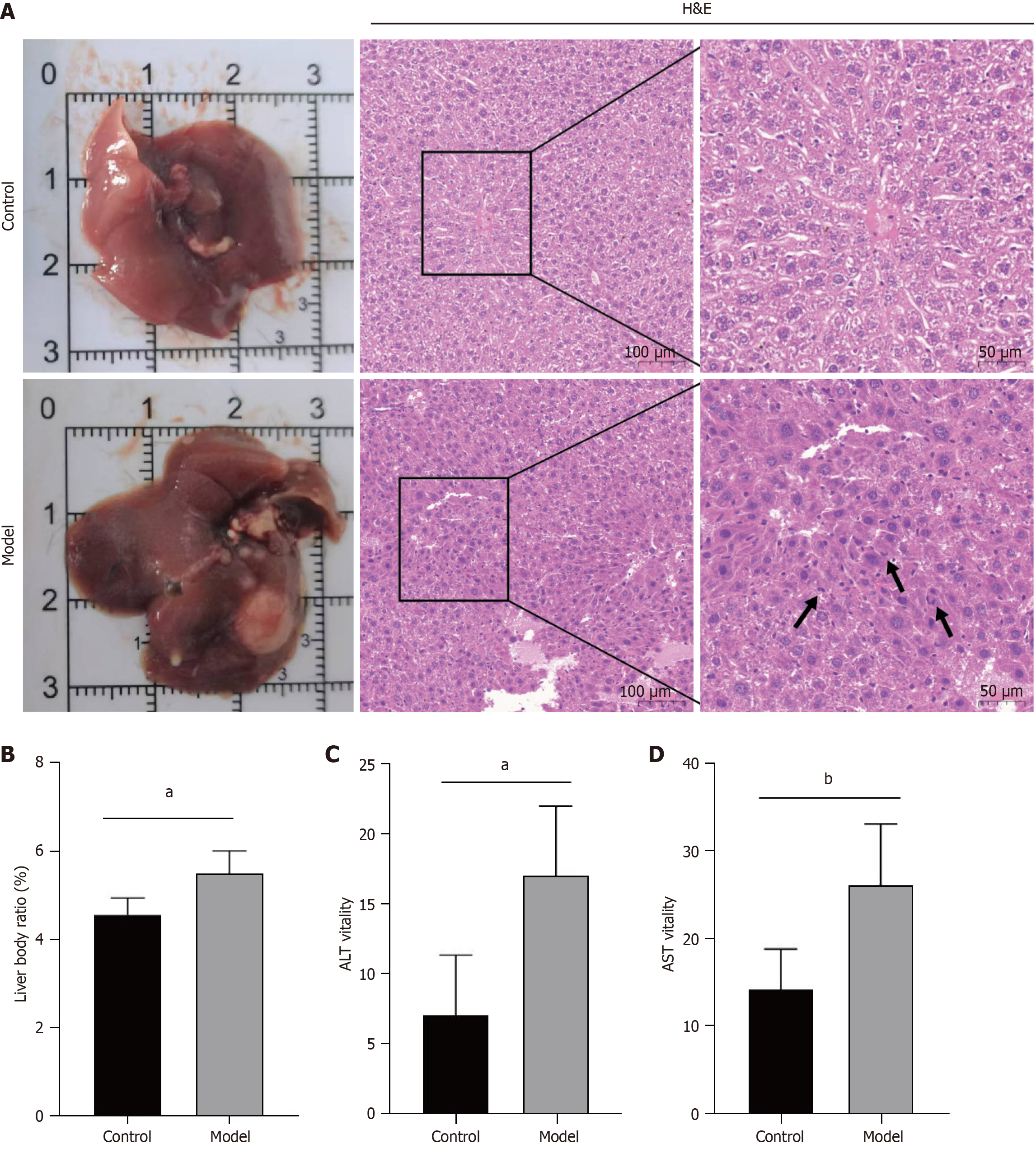
Figure 5 Evaluation of hepatocellular carcinoma model induction efficacy.
A: Appearance and hemotoxylin and eosin staining of liver lobules in the control and model groups. Abnormal mitosis of hepatocellular carcinoma cells is indicated by black arrows; magnification, 200 ×; B: Liver body ratios for the control and model mice; C: Alanine aminotransferase for the control and model mice; D: Aspartate aminotransferase for the control and model mice. H&E: Hemotoxylin and eosin; ALT: Alanine aminotransferase; AST: Aspartate aminotransferase. aP < 0.001, bP < 0.01.
- Citation: Guo W, Wang MX, Mu T, Xu K, Jiang ZD, Wan XH, Li JW, Yi YX. Reduction in the hepatocellular carcinoma antioncogene SLC39A14 during the malignant progression of hepatocellular carcinoma. World J Gastrointest Surg 2025; 17(10): 111454
- URL: https://www.wjgnet.com/1948-9366/full/v17/i10/111454.htm
- DOI: https://dx.doi.org/10.4240/wjgs.v17.i10.111454