
















©The Author(s) 2025.
World J Gastrointest Surg. Oct 27, 2025; 17(10): 109920
Published online Oct 27, 2025. doi: 10.4240/wjgs.v17.i10.109920
Published online Oct 27, 2025. doi: 10.4240/wjgs.v17.i10.109920
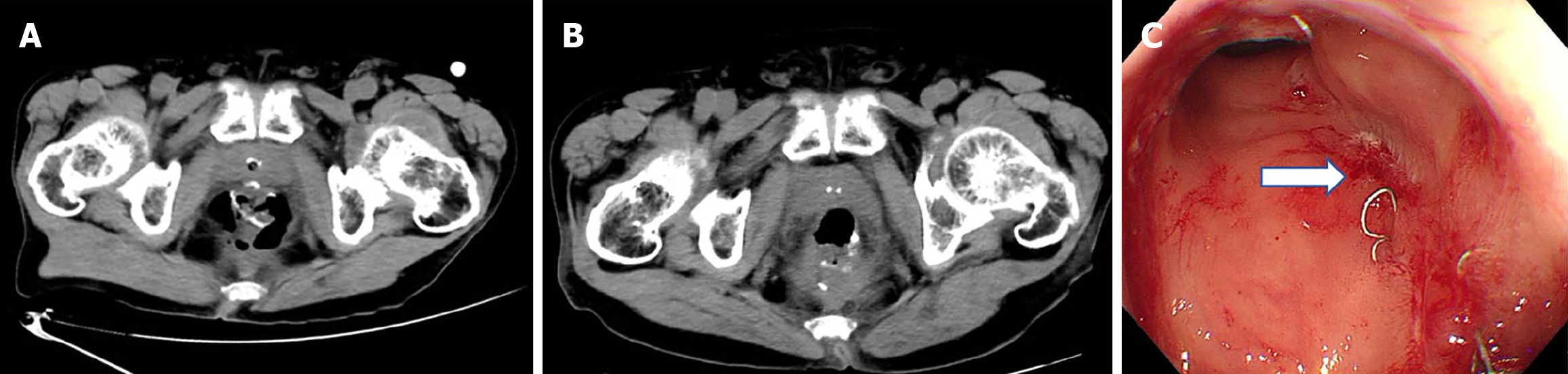
Figure 3 Case 2.
A: Abdominal computed tomography scan revealed a discontinuous anastomosis and the formation of an anastomotic leak; B: Subsequently after transanal opening of the intersphincteric space, an abdominal computed tomography scan showed that the anastomosis was continuous with no obvious gap; C: The colonoscopy confirmed that the anastomotic defect was healed. Arrows: Healing site of the anastomotic fistula opening.
- Citation: Li H, Huang HB, Xiang T, Yang L, Furnée EJB, Sun G, Chen WB. Transanal intersphincteric approach combined with Kangfuxin enema for treating anastomotic leakage after low anterior resection: Three case reports. World J Gastrointest Surg 2025; 17(10): 109920
- URL: https://www.wjgnet.com/1948-9366/full/v17/i10/109920.htm
- DOI: https://dx.doi.org/10.4240/wjgs.v17.i10.109920