
















©The Author(s) 2025.
World J Gastrointest Surg. Jan 27, 2025; 17(1): 100278
Published online Jan 27, 2025. doi: 10.4240/wjgs.v17.i1.100278
Published online Jan 27, 2025. doi: 10.4240/wjgs.v17.i1.100278
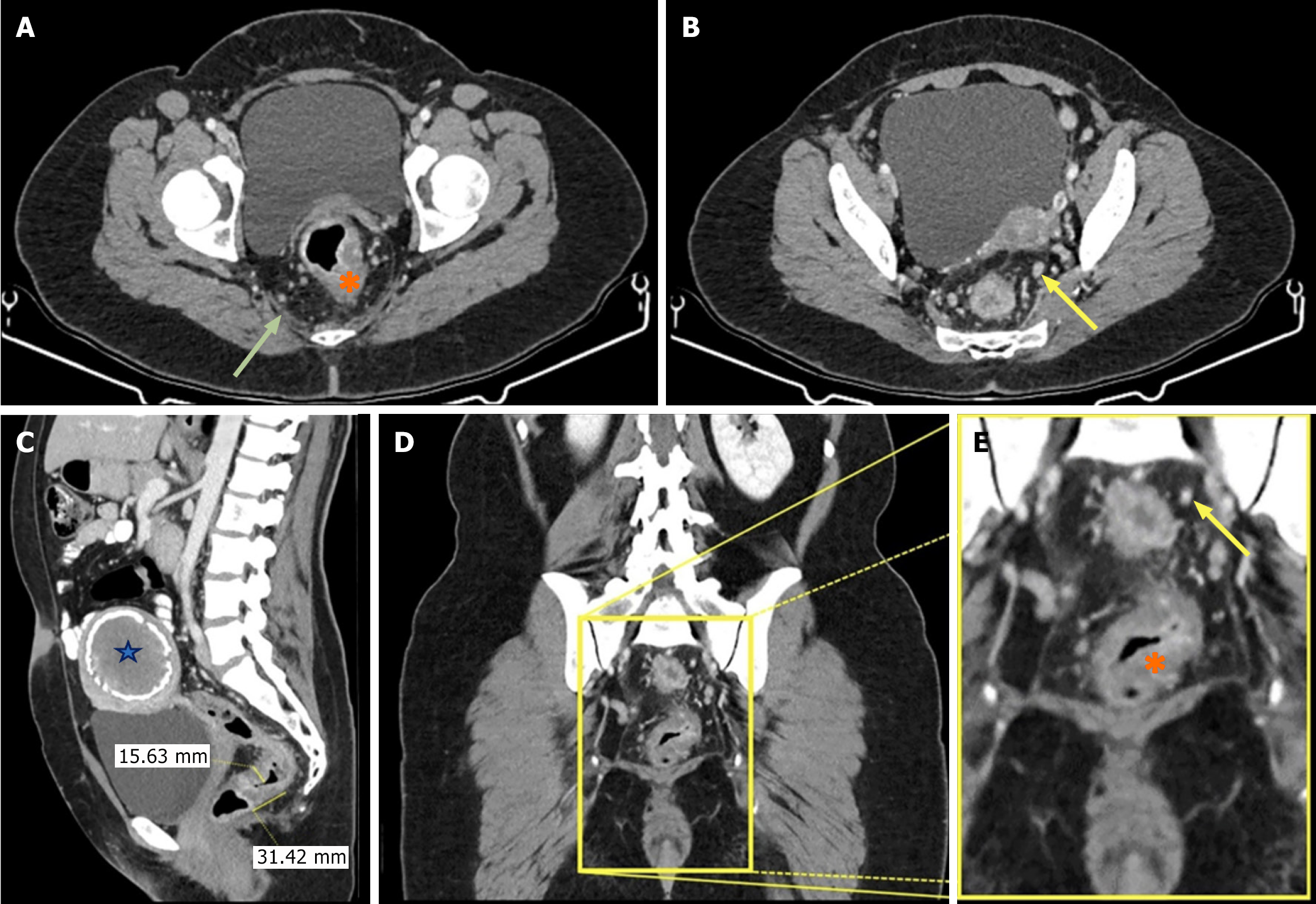
Figure 1 Contrast-enhanced pelvic computed tomography images of a 46-year-old female patient.
A: Lesion suggestive of malignancy on the rectal wall, ameboma (orange asterisk), increased density in the pararectal mesenteric tissue (green arrow); B: Only one of the lymph nodes in the pararectal region increased in number (yellow arrow); C: Sagittal image shows increased thickness of the rectal wall and calcified myoma (blue star) in the uterus; D and E: It is the magnified image of coronal plane image (D). This magnified image shows increased thickness of the rectal wall (orange asterisk) and lymph nodes in the coronal section (yellow arrow).
- Citation: Memis KB, Celik AS, Aydin S, Kantarci M. Rectal ameboma: A new entity in the differential diagnosis of rectal cancer. World J Gastrointest Surg 2025; 17(1): 100278
- URL: https://www.wjgnet.com/1948-9366/full/v17/i1/100278.htm
- DOI: https://dx.doi.org/10.4240/wjgs.v17.i1.100278