Copyright
©The Author(s) 2024.
World J Gastrointest Surg. Feb 27, 2024; 16(2): 609-615
Published online Feb 27, 2024. doi: 10.4240/wjgs.v16.i2.609
Published online Feb 27, 2024. doi: 10.4240/wjgs.v16.i2.609
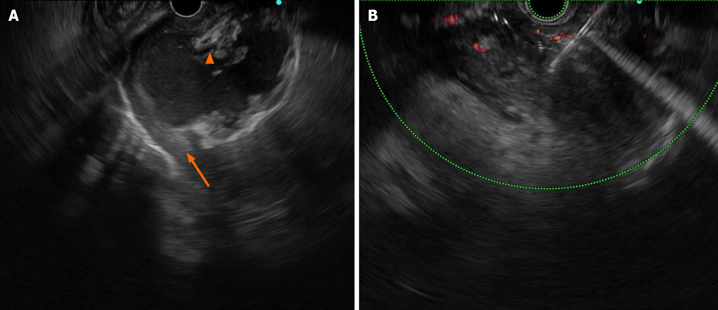
Figure 2 Endoscopic ultrasound-guided aspiration and lavage.
A: The size of the cyst cavity was 56 mm × 36 mm, with necrotic debris (orange triangle), and the necrotic collection was not walled-off (orange arrows); B: The cystic cavity almost disappeared after intervention.
- Citation: Zhang HY, He CC. Early endoscopic management of an infected acute necrotic collection misdiagnosed as a pancreatic pseudocyst: A case report. World J Gastrointest Surg 2024; 16(2): 609-615
- URL: https://www.wjgnet.com/1948-9366/full/v16/i2/609.htm
- DOI: https://dx.doi.org/10.4240/wjgs.v16.i2.609


