
















©The Author(s) 2024.
World J Gastrointest Surg. Oct 27, 2024; 16(10): 3312-3320
Published online Oct 27, 2024. doi: 10.4240/wjgs.v16.i10.3312
Published online Oct 27, 2024. doi: 10.4240/wjgs.v16.i10.3312
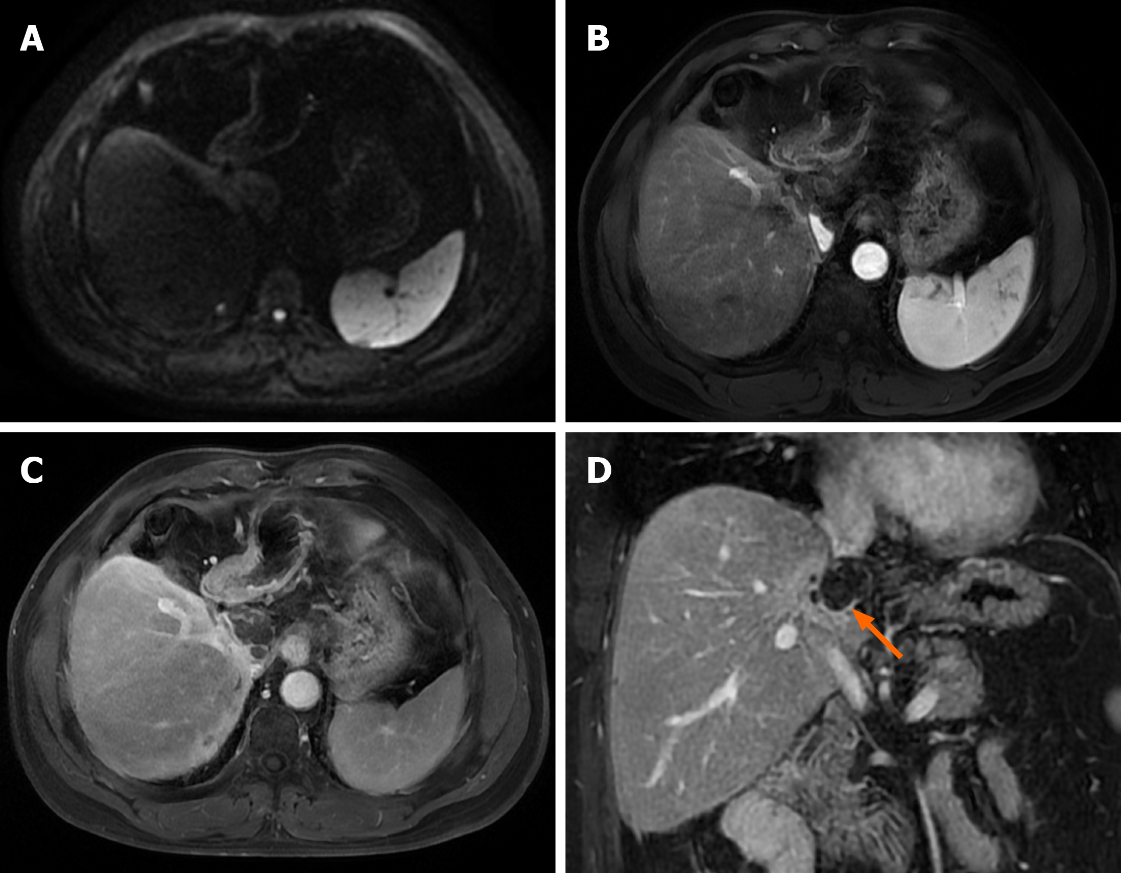
Figure 4 Post-operative magnetic resonance imaging of case 2 (6 months).
A: Diffusion-weighted phase revealed no obvious tumor activity in the residual liver; B and C: T1-weighted enhanced magnetic resonance imaging showed extensive tumor necrosis in the caudate lobe, and local slight enhancement around the tumor showing high signal in arterial phase and venous phase; D: Coronal delayed phase confirmed extensive tumor necrosis in the caudate lobe with marginal local slight enhancement (arrow) and no tumor activity in the remaining right liver.
- Citation: Zhu YB, Qin JY, Zhang TT, Zhang WJ, Ling Q. Reassessment of palliative surgery in conversion therapy of previously unresectable hepatocellular carcinoma: Two case reports and review of literature. World J Gastrointest Surg 2024; 16(10): 3312-3320
- URL: https://www.wjgnet.com/1948-9366/full/v16/i10/3312.htm
- DOI: https://dx.doi.org/10.4240/wjgs.v16.i10.3312