Copyright
©The Author(s) 2024.
World J Gastrointest Surg. Jan 27, 2024; 16(1): 239-247
Published online Jan 27, 2024. doi: 10.4240/wjgs.v16.i1.239
Published online Jan 27, 2024. doi: 10.4240/wjgs.v16.i1.239
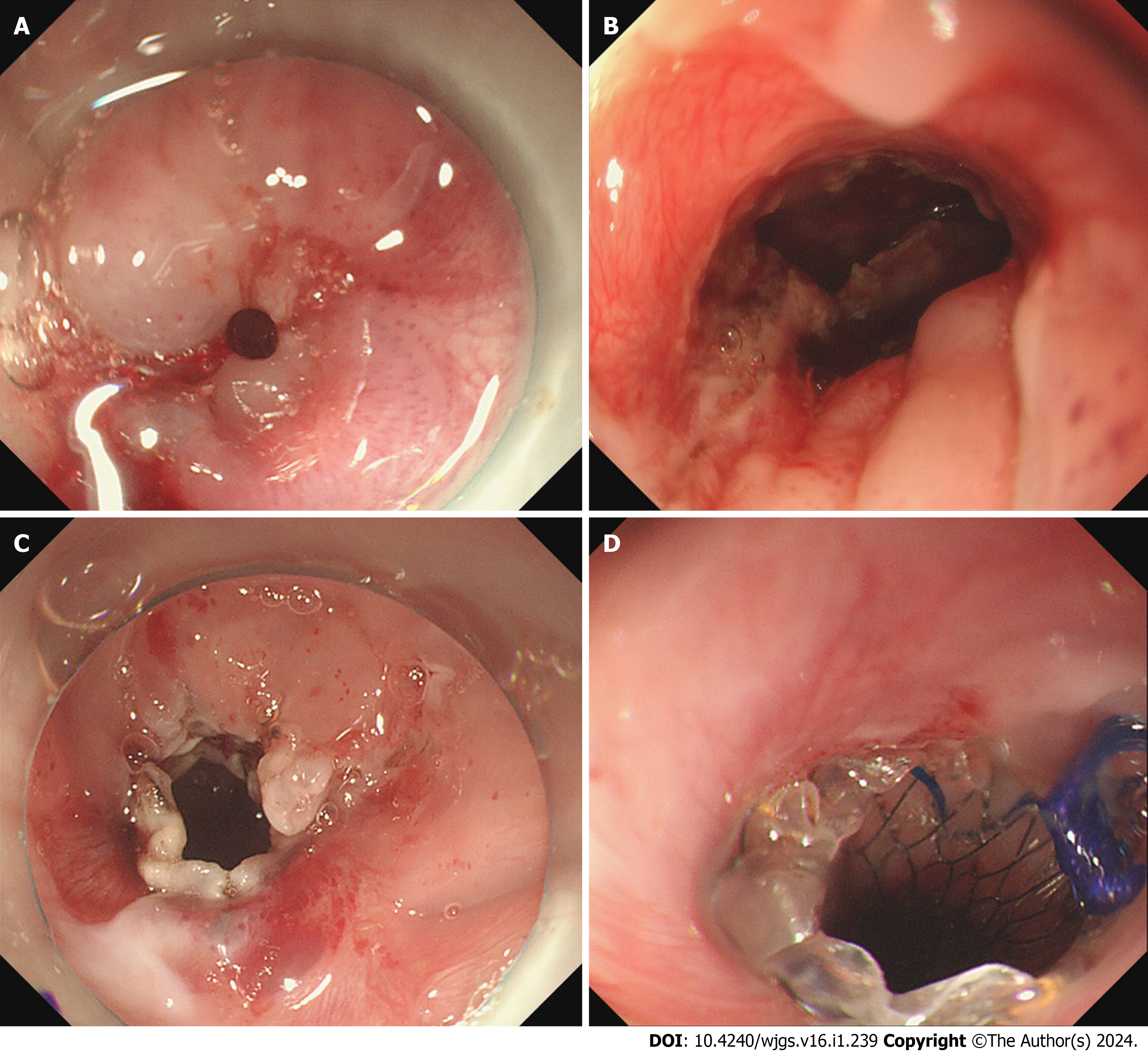
Figure 4 The endoscopic incision method and esophageal stent placement performed at admission.
A: The esophageal stenosis part which made the endoscope unable to enter; B: There were fistulas in the patient's esophagus; C: The esophageal lumen increased significantly after treatment by the endoscopic incision method; D: The upper end of the stent after placement.
- Citation: Fang JH, Li WM, He CH, Wu JL, Guo Y, Lai ZC, Li GD. Endoscopic treatment of extreme esophageal stenosis complicated with esophagotracheal fistula: A case report. World J Gastrointest Surg 2024; 16(1): 239-247
- URL: https://www.wjgnet.com/1948-9366/full/v16/i1/239.htm
- DOI: https://dx.doi.org/10.4240/wjgs.v16.i1.239


