Copyright
©The Author(s) 2023.
World J Gastrointest Surg. Aug 27, 2023; 15(8): 1831-1837
Published online Aug 27, 2023. doi: 10.4240/wjgs.v15.i8.1831
Published online Aug 27, 2023. doi: 10.4240/wjgs.v15.i8.1831
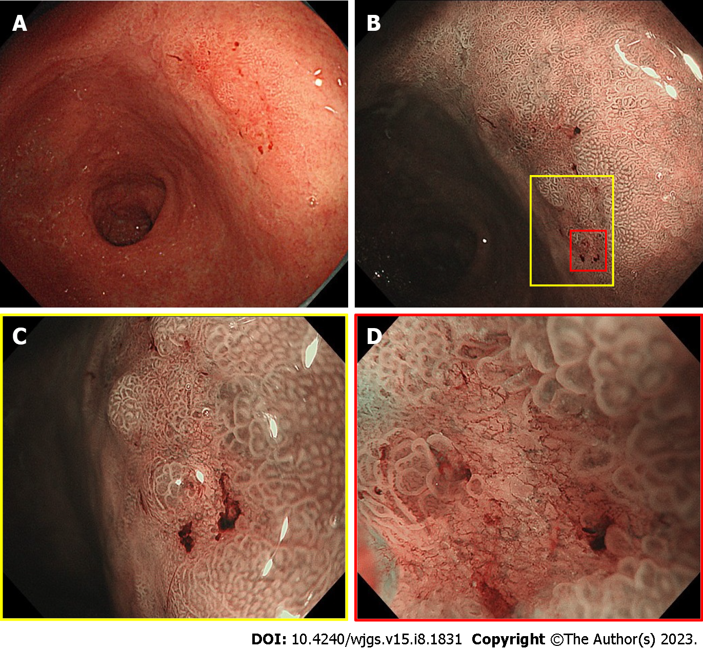
Figure 2 Endoscopic view of the 10-mm flat-depressed lesion on the posterior wall of the gastric antrum.
A: Distant view of the lesion under white-light imaging; B: Non-expansion view of the lesion using magnifying endoscopy with narrow-band imaging; C: Weak-expansion view of the lesion using magnifying endoscopy with narrow-band imaging; D: Strong-expansion view of the lesion using magnifying endoscopy with narrow-band imaging.
- Citation: Ito R, Miwa K, Matano Y. Outpatient hybrid endoscopic submucosal dissection with SOUTEN for early gastric cancer, followed by endoscopic suturing of the mucosal defect: A case report. World J Gastrointest Surg 2023; 15(8): 1831-1837
- URL: https://www.wjgnet.com/1948-9366/full/v15/i8/1831.htm
- DOI: https://dx.doi.org/10.4240/wjgs.v15.i8.1831


