Copyright
©The Author(s) 2023.
World J Gastrointest Surg. Jul 27, 2023; 15(7): 1331-1339
Published online Jul 27, 2023. doi: 10.4240/wjgs.v15.i7.1331
Published online Jul 27, 2023. doi: 10.4240/wjgs.v15.i7.1331
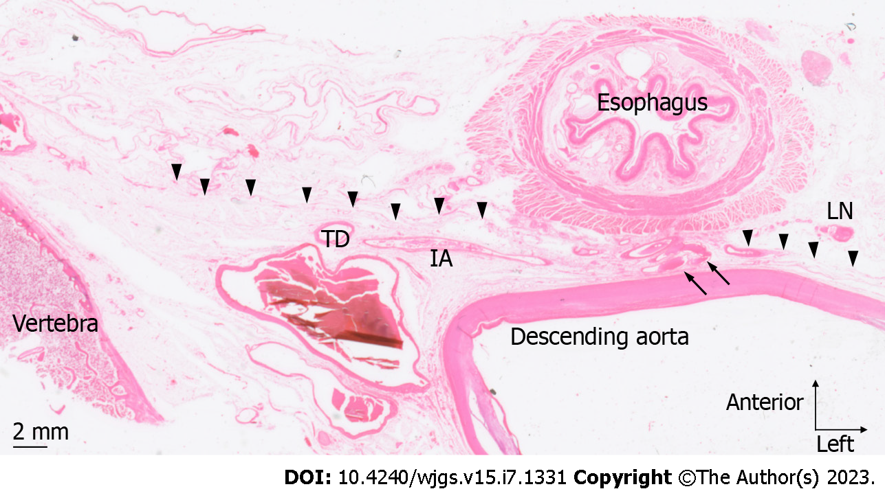
Figure 3 Artery penetrating fascia B.
Cadaver 5, HE-stained section 9 mm cranial to the esophageal hiatus. An artery (arrow) branching from the anterior surface of the descending aorta penetrates fascia B (arrowhead) and runs toward the esophageal wall. No lymph nodes were observed around this artery. TD: Thoracic duct; IA: Intercostal artery; LN: Lymph node.
- Citation: Saito T, Muro S, Fujiwara H, Umebayashi Y, Sato Y, Tokunaga M, Akita K, Kinugasa Y. Histological study of the structural layers around the esophagus in the lower mediastinum. World J Gastrointest Surg 2023; 15(7): 1331-1339
- URL: https://www.wjgnet.com/1948-9366/full/v15/i7/1331.htm
- DOI: https://dx.doi.org/10.4240/wjgs.v15.i7.1331


