Copyright
©The Author(s) 2023.
World J Gastrointest Surg. Jun 27, 2023; 15(6): 1116-1124
Published online Jun 27, 2023. doi: 10.4240/wjgs.v15.i6.1116
Published online Jun 27, 2023. doi: 10.4240/wjgs.v15.i6.1116
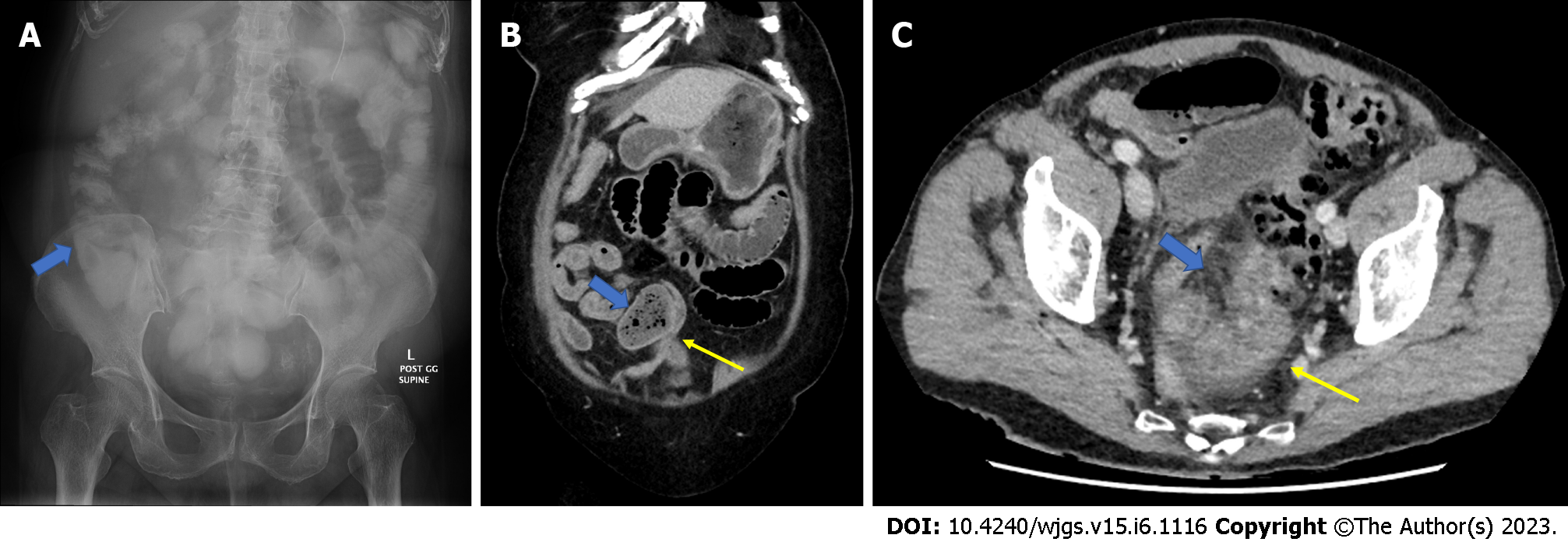
Figure 1 Images.
A: Abdominal X-ray showing the presence of water soluble contrast medium in the large colon; B: Coronal slice of the computed tomography (CT) scan showing small bowel faecal sign (blue arrow) and transition point (yellow arrow) in adhesive small bowel obstruction; C: Axial slice of CT scan showing a segment of small bowel thickening/reduced wall enhancement (yellow arrow) with mesenteric stranding (blue arrow) in the presence of small bowel obstruction.
- Citation: Ng ZQ, Hsu V, Tee WWH, Tan JH, Wijesuriya R. Predictors for success of non-operative management of adhesive small bowel obstruction. World J Gastrointest Surg 2023; 15(6): 1116-1124
- URL: https://www.wjgnet.com/1948-9366/full/v15/i6/1116.htm
- DOI: https://dx.doi.org/10.4240/wjgs.v15.i6.1116


