Copyright
©The Author(s) 2023.
World J Gastrointest Surg. May 27, 2023; 15(5): 992-999
Published online May 27, 2023. doi: 10.4240/wjgs.v15.i5.992
Published online May 27, 2023. doi: 10.4240/wjgs.v15.i5.992
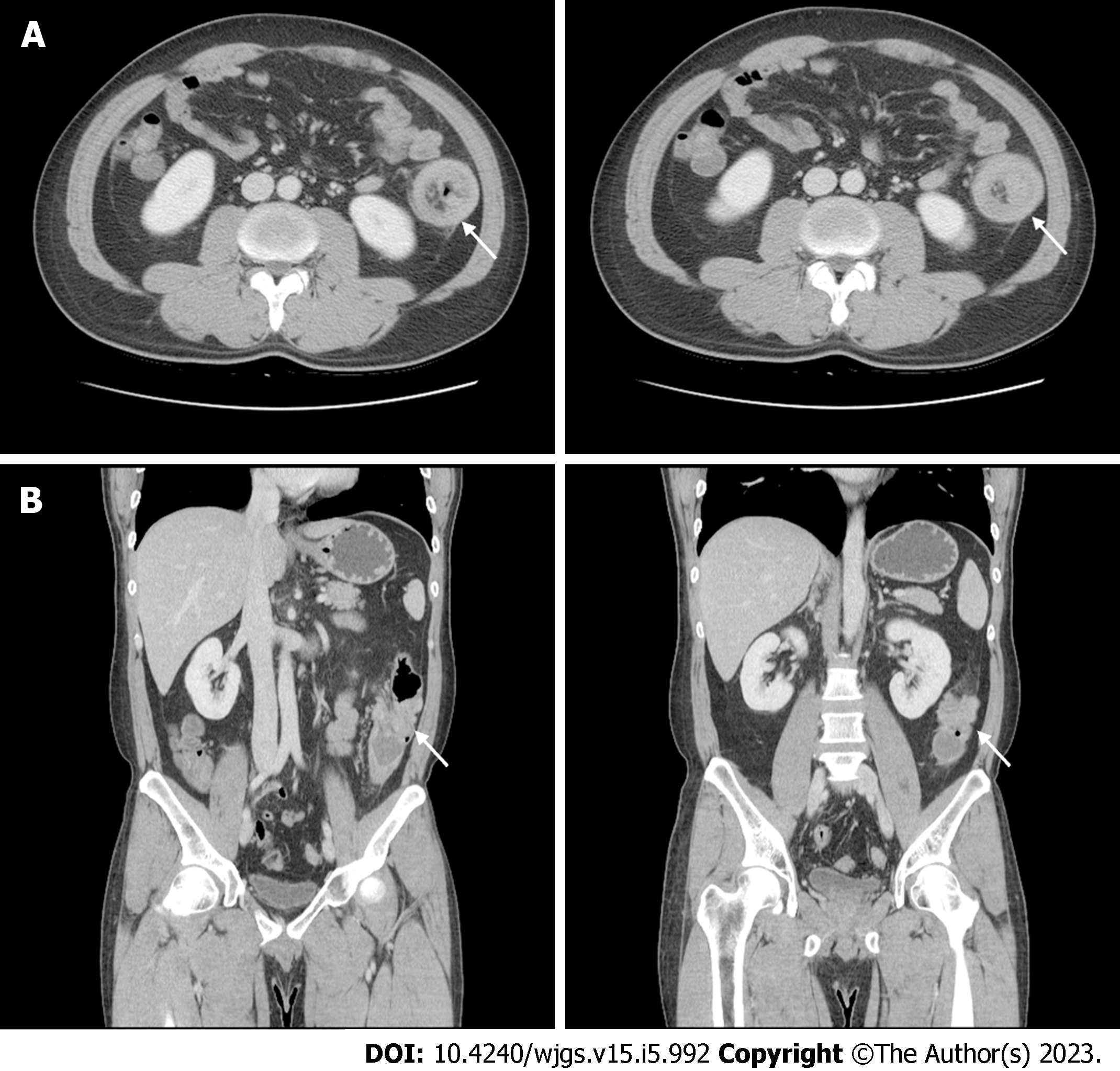
Figure 2 Contrast-enhanced abdominal computed tomography.
Computed tomography shows the lead point (white arrow) of the intussusception in the descending colon due to an approximately 3-cm cystic mass. A: Axial view; B: Coronal view.
- Citation: Lee SH, Bae SH, Lee SC, Ahn TS, Kim Z, Jung HI. Curative resection of leiomyosarcoma of the descending colon with metachronous liver metastasis: A case report. World J Gastrointest Surg 2023; 15(5): 992-999
- URL: https://www.wjgnet.com/1948-9366/full/v15/i5/992.htm
- DOI: https://dx.doi.org/10.4240/wjgs.v15.i5.992


