Copyright
©The Author(s) 2023.
World J Gastrointest Surg. Apr 27, 2023; 15(4): 600-620
Published online Apr 27, 2023. doi: 10.4240/wjgs.v15.i4.600
Published online Apr 27, 2023. doi: 10.4240/wjgs.v15.i4.600
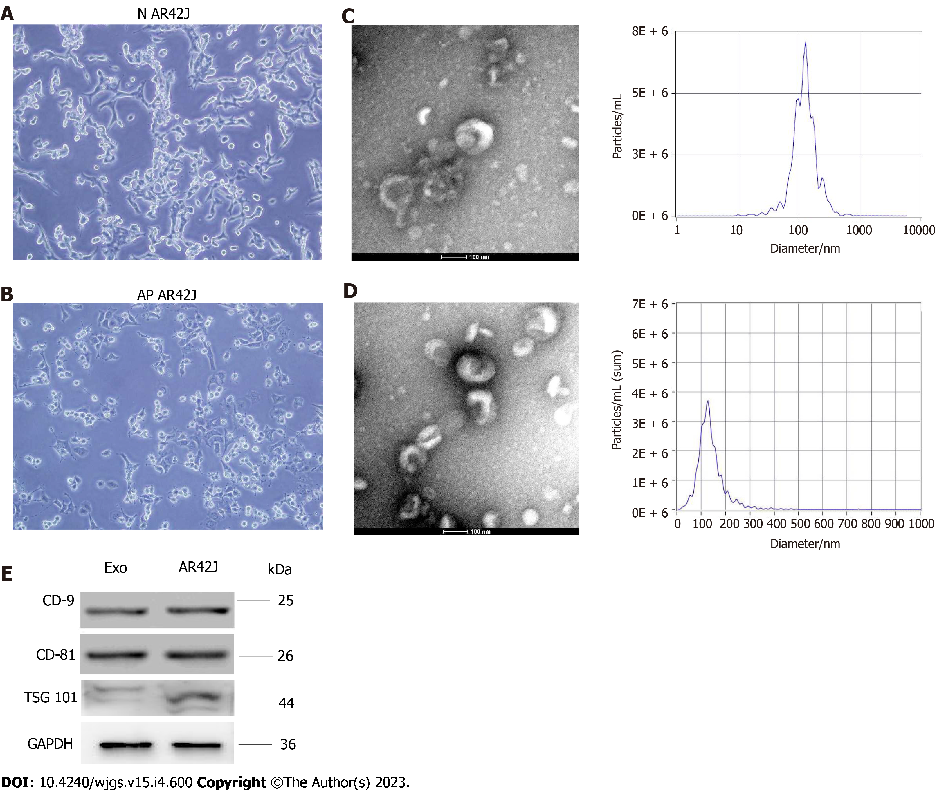
Figure 1 Isolation and characteristics of exosomes derived from normal acinous cell AR42J and active AR42J.
A: Morphology of AR42J cell line in the inactive state; B: Morphology of AR42J cell line in the activated state; C: Non-activated AR42J exosome morphology shown by transmission electron microscope (TEM) at 100nm scale. The diameter of exosomes derived from AR42J in the inactive state ranged from 68.3–181.9 nm, with an average of 121.4 nm, and the concentration of exosomes was 3.3 × 210 particles /mL; D: Morphology of AR42J exosomes in activated state as shown by TEM at 100 nm scale. The diameter of exosomes derived from AR42J in the activated state ranged from 82.1–197.5 nm, with an average of 132.8 nm, and the concentration of exosomes was 6.23 × 210 particles /mL; E: The positive expression of exosome marker, which include CD9, CD81 and TSG101, was confirmed by Western blot. AP: Acute pancreatitis; N: Normal.
- Citation: Zheng Z, Cao F, Ding YX, Lu JD, Fu YQ, Liu L, Guo YL, Liu S, Sun HC, Cui YQ, Li F. Acinous cell AR42J-derived exosome miR125b-5p promotes acute pancreatitis exacerbation by inhibiting M2 macrophage polarization via PI3K/AKT signaling pathway. World J Gastrointest Surg 2023; 15(4): 600-620
- URL: https://www.wjgnet.com/1948-9366/full/v15/i4/600.htm
- DOI: https://dx.doi.org/10.4240/wjgs.v15.i4.600


