
















©The Author(s) 2023.
World J Gastrointest Surg. Mar 27, 2023; 15(3): 488-494
Published online Mar 27, 2023. doi: 10.4240/wjgs.v15.i3.488
Published online Mar 27, 2023. doi: 10.4240/wjgs.v15.i3.488
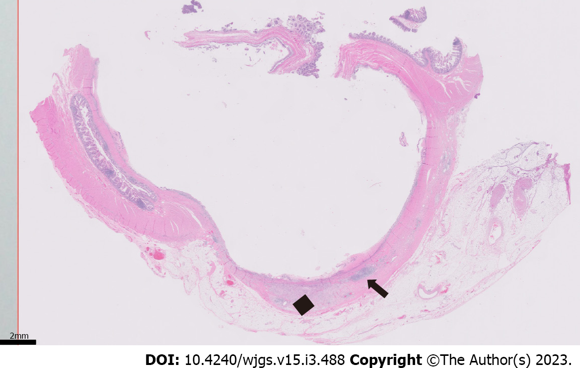
Figure 4 Low-magnification section of the specimen.
Microscopic (0.5 × objective magnification) cross section of the cystically dilated small intestine with mucosal ulceration (diffusely involving the luminal surface) and chronic inflammation (arrow), fibrosis, and xanthogranulomatous inflammation (black diamond) of the underlying wall.
- Citation: Wang W, Korah M, Bessoff KE, Shen J, Forrester JD. Xanthogranulomatous inflammation requiring small bowel anastomosis revision: A case report. World J Gastrointest Surg 2023; 15(3): 488-494
- URL: https://www.wjgnet.com/1948-9366/full/v15/i3/488.htm
- DOI: https://dx.doi.org/10.4240/wjgs.v15.i3.488