Copyright
©The Author(s) 2023.
World J Gastrointest Surg. Dec 27, 2023; 15(12): 2693-2708
Published online Dec 27, 2023. doi: 10.4240/wjgs.v15.i12.2693
Published online Dec 27, 2023. doi: 10.4240/wjgs.v15.i12.2693
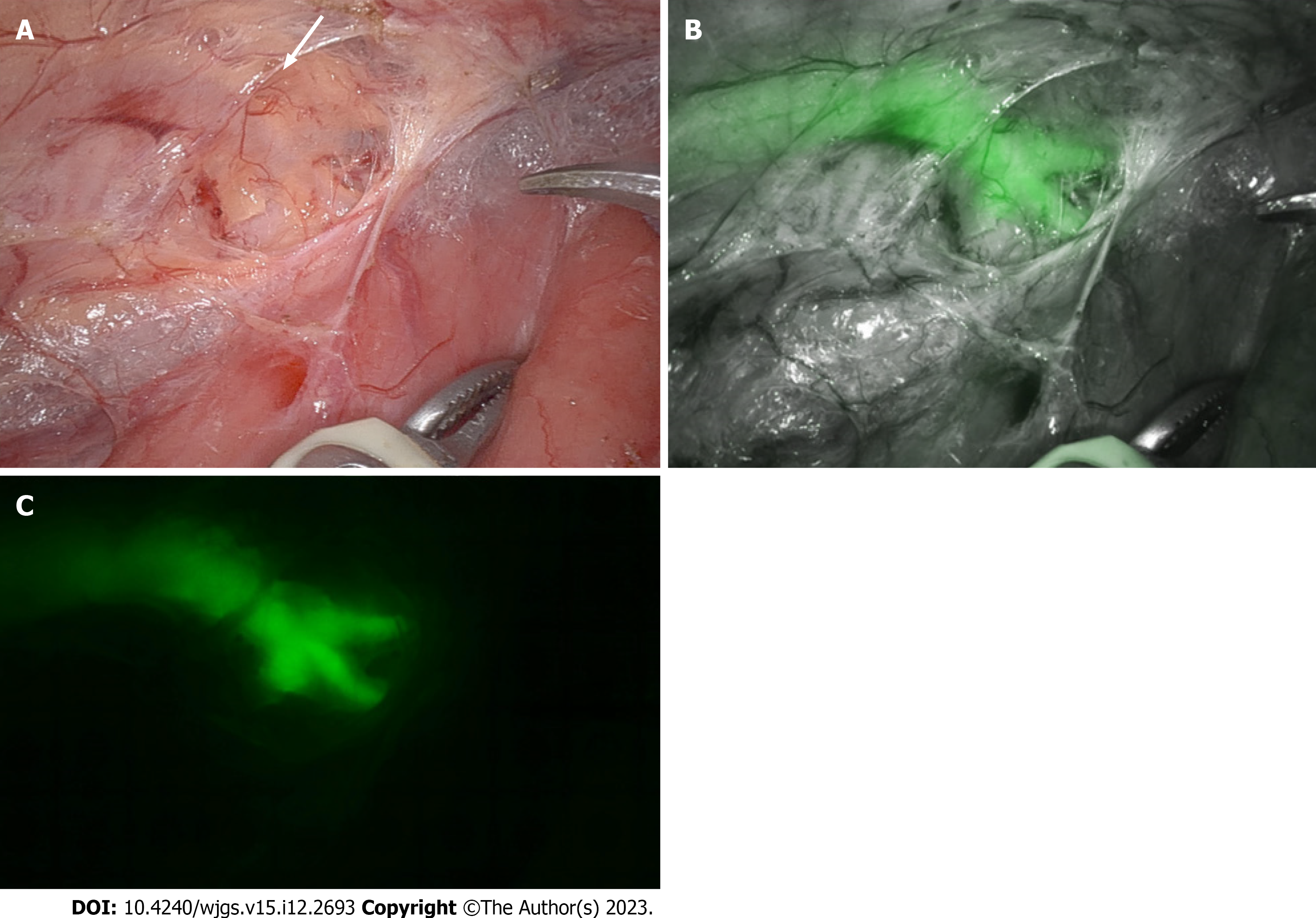
Figure 5 Visualization of the thoracic duct.
A and B: Visualization of the thoracic duct during robotic esophagectomy in normal mode (A) is enhanced by indocyanine green fluorescence (B); C: The branching pattern of the thoracic duct is well visualized in fluorescence mode.
- Citation: Kalayarasan R, Chandrasekar M, Sai Krishna P, Shanmugam D. Indocyanine green fluorescence in gastrointestinal surgery: Appraisal of current evidence. World J Gastrointest Surg 2023; 15(12): 2693-2708
- URL: https://www.wjgnet.com/1948-9366/full/v15/i12/2693.htm
- DOI: https://dx.doi.org/10.4240/wjgs.v15.i12.2693


