Copyright
©The Author(s) 2022.
World J Gastrointest Surg. Apr 27, 2022; 14(4): 341-351
Published online Apr 27, 2022. doi: 10.4240/wjgs.v14.i4.341
Published online Apr 27, 2022. doi: 10.4240/wjgs.v14.i4.341
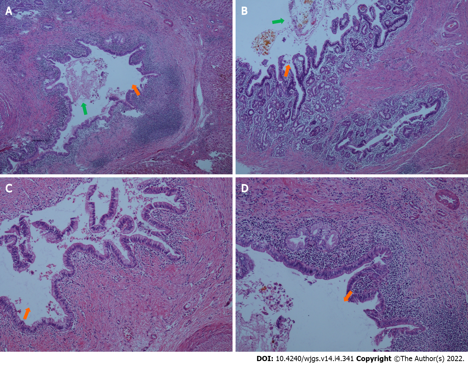
Figure 6 Histopathological findings of the resected liver (intrahepatic duct).
A and B: Representative specimens showing intrahepatic duct dilatation (orange arrows) with stones (green arrows) and inflammatory cell infiltration in bile duct walls (hematoxylin and eosin staining; 40 ×); C and D: Representative specimens showing intrahepatic duct dilatation (orange arrows) with stones and inflammatory cell infiltration in bile duct walls (hematoxylin and eosin staining; 100 ×).
- Citation: Fan WJ, Zou XJ. Subacute liver and respiratory failure after segmental hepatectomy for complicated hepatolithiasis with secondary biliary cirrhosis: A case report. World J Gastrointest Surg 2022; 14(4): 341-351
- URL: https://www.wjgnet.com/1948-9366/full/v14/i4/341.htm
- DOI: https://dx.doi.org/10.4240/wjgs.v14.i4.341


