
















©The Author(s) 2022.
World J Gastrointest Surg. Apr 27, 2022; 14(4): 341-351
Published online Apr 27, 2022. doi: 10.4240/wjgs.v14.i4.341
Published online Apr 27, 2022. doi: 10.4240/wjgs.v14.i4.341
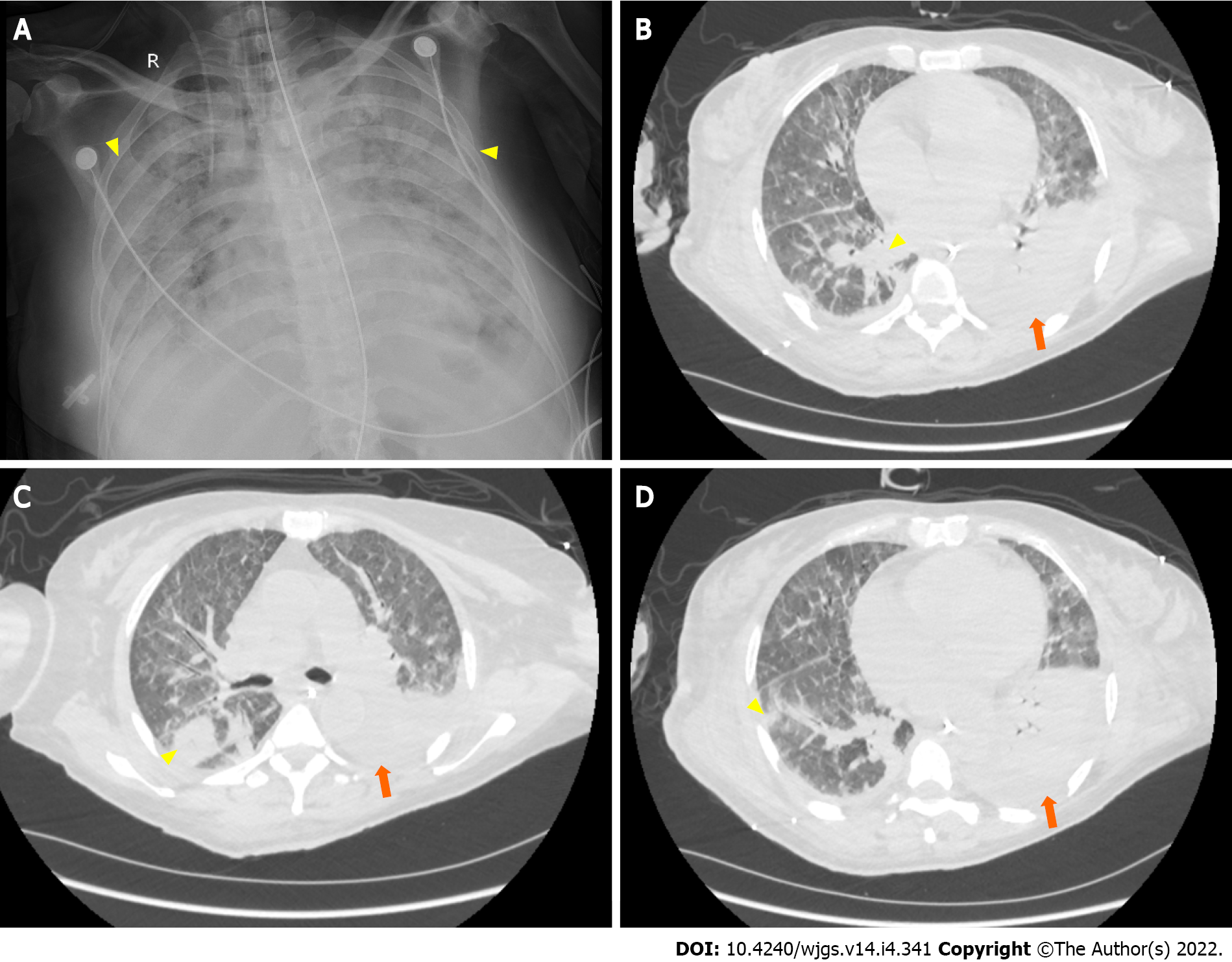
Figure 5 Postoperative pulmonary imaging.
A: Bedside chest X-ray (August 14, 2021) showing bilateral pulmonary diffuse patchy high-density shadows (yellow arrows) and bilateral pleural effusion, indicating bilateral pulmonary infection; B-D: Representative chest computed tomography (August 20, 2021) images showing bilateral pulmonary nodular and patchy shadows (yellow arrows) and left pulmonary atelectasis (orange arrows), indicating pulmonary infection.
- Citation: Fan WJ, Zou XJ. Subacute liver and respiratory failure after segmental hepatectomy for complicated hepatolithiasis with secondary biliary cirrhosis: A case report. World J Gastrointest Surg 2022; 14(4): 341-351
- URL: https://www.wjgnet.com/1948-9366/full/v14/i4/341.htm
- DOI: https://dx.doi.org/10.4240/wjgs.v14.i4.341