Copyright
©The Author(s) 2022.
World J Gastrointest Surg. Nov 27, 2022; 14(11): 1310-1319
Published online Nov 27, 2022. doi: 10.4240/wjgs.v14.i11.1310
Published online Nov 27, 2022. doi: 10.4240/wjgs.v14.i11.1310
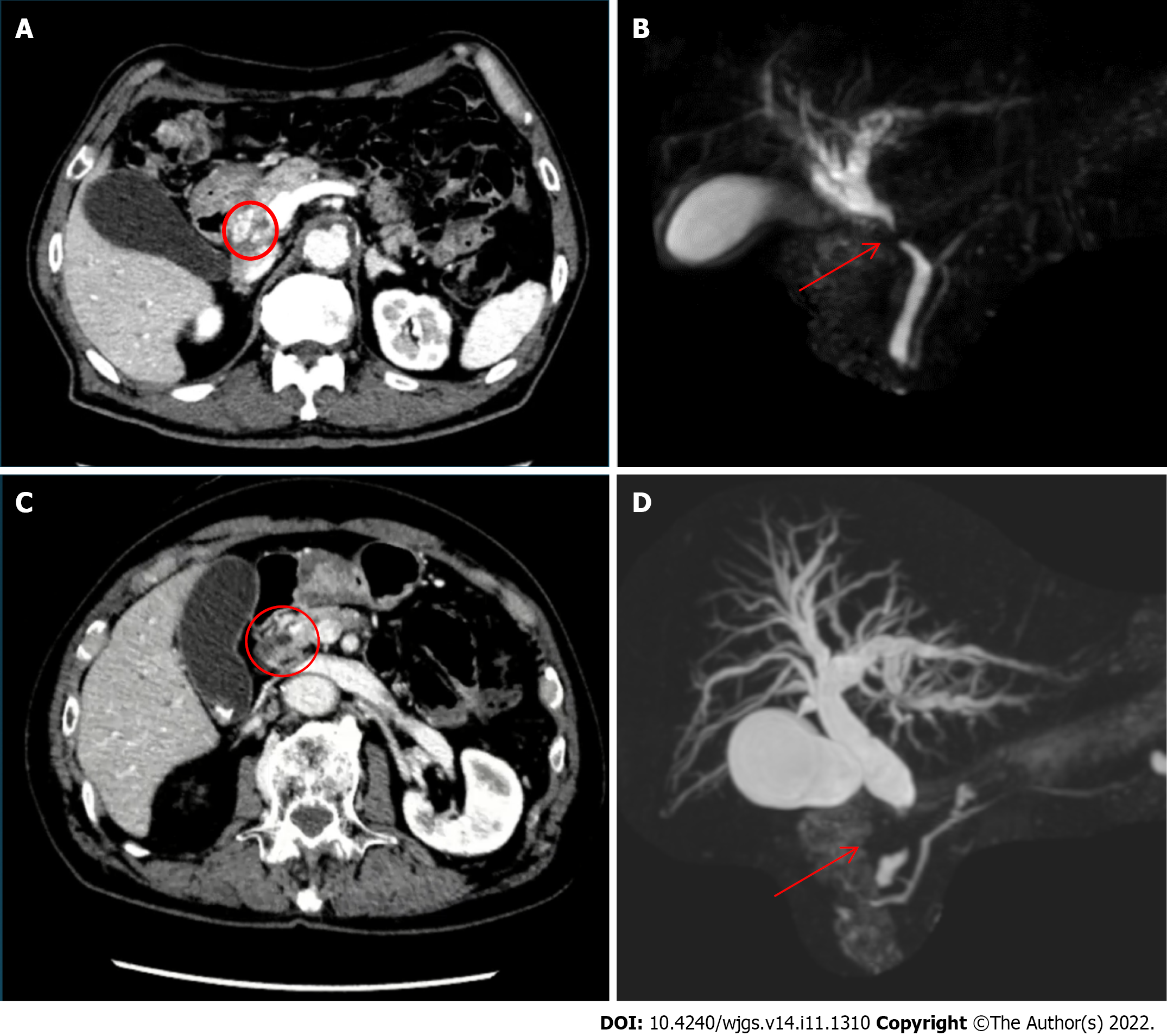
Figure 1 Imaging examinations.
A: Dynamic computed tomography (case 1) showing common bile duct wall thickening at the ductal confluence (circle) and mild dilatation of the intrahepatic bile duct; B: Magnetic resonance cholangiopancreatography showing stricture of common bile duct (arrow) and dilatation of the intrahepatic bile duct; C: Dynamic computed tomography (case 2) showing bile duct wall thickening at the upper pancreatic margin level (circle) with dilatation of the entire bile duct system without pancreatic duct dilatation; D: Magnetic resonance cholangiopancreatography showing stenosis of the distal common hepatic duct (arrow) and dilatation of the intrahepatic bile duct.
- Citation: Colella M, Mishima K, Wakabayashi T, Fujiyama Y, Al-Omari MA, Wakabayashi G. Preoperative blood circulation modification prior to pancreaticoduodenectomy in patients with celiac trunk occlusion: Two case reports. World J Gastrointest Surg 2022; 14(11): 1310-1319
- URL: https://www.wjgnet.com/1948-9366/full/v14/i11/1310.htm
- DOI: https://dx.doi.org/10.4240/wjgs.v14.i11.1310


