Copyright
©The Author(s) 2022.
World J Gastrointest Surg. Oct 27, 2022; 14(10): 1169-1178
Published online Oct 27, 2022. doi: 10.4240/wjgs.v14.i10.1169
Published online Oct 27, 2022. doi: 10.4240/wjgs.v14.i10.1169
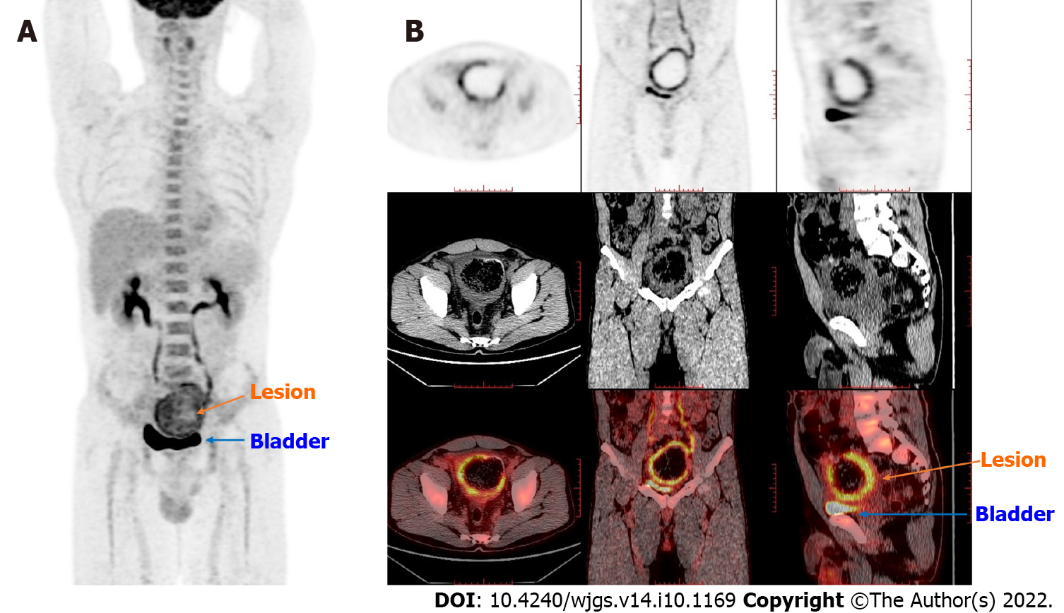
Figure 2 Positron emission tomography/computed tomography findings.
The thick-walled cystic structure above the bladder could be seen, the radioactive uptake was significantly increased, and the maximum standard uptake value reached 6.5. The diagnosis was considered inflammatory or a neoplastic lesion on the sigmoid colon adherent to the bladder. A: Coronal whole-body imaging showing high uptake values at the lesion site; B: Detailed imaging pictures of the lesion, including horizontal, sagittal and coronal.
- Citation: Zhan WL, Liu L, Jiang W, He FX, Qu HT, Cao ZX, Xu XS. Immunoglobulin G4-related disease in the sigmoid colon in patient with severe colonic fibrosis and obstruction: A case report. World J Gastrointest Surg 2022; 14(10): 1169-1178
- URL: https://www.wjgnet.com/1948-9366/full/v14/i10/1169.htm
- DOI: https://dx.doi.org/10.4240/wjgs.v14.i10.1169


