Copyright
©The Author(s) 2022.
World J Gastrointest Surg. Oct 27, 2022; 14(10): 1161-1168
Published online Oct 27, 2022. doi: 10.4240/wjgs.v14.i10.1161
Published online Oct 27, 2022. doi: 10.4240/wjgs.v14.i10.1161
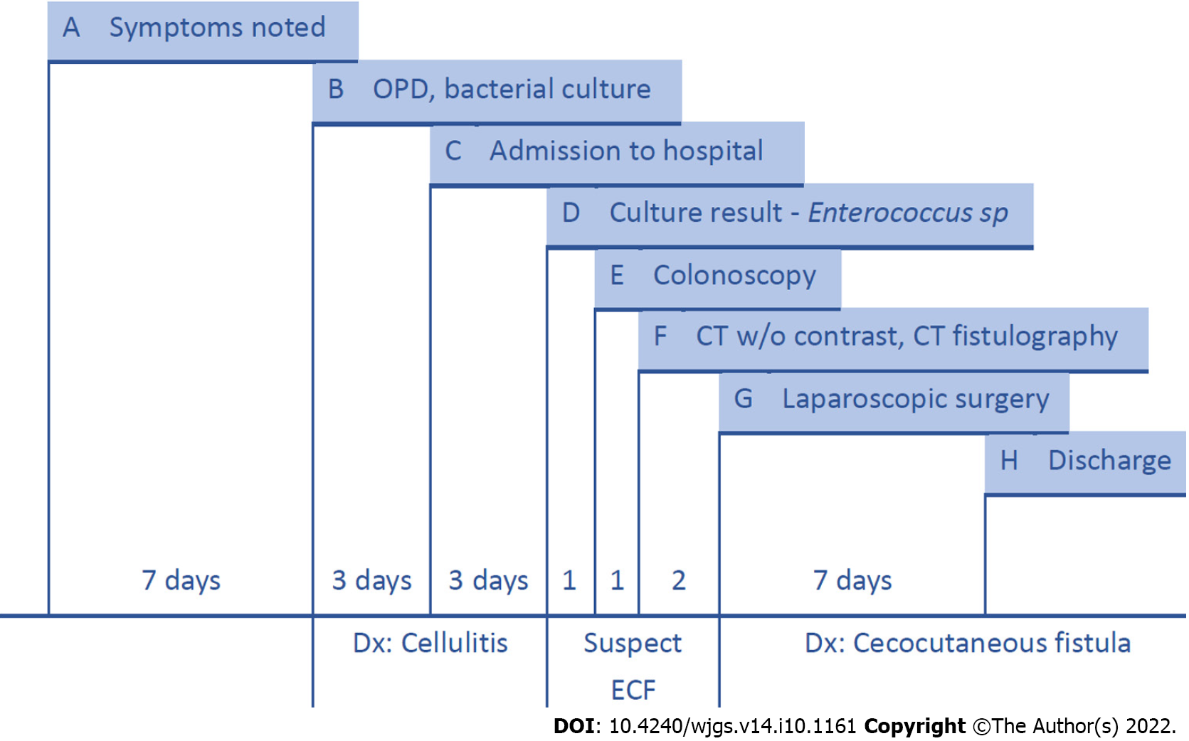
Figure 5 Timeline.
A: Redness and swelling over the right lower quadrant abdominal wall were noted; B: The patient came to the outpatient department for help; discharge from the carbuncle abscess was collected for bacterial culture. Blood analysis was performed; C: The patient was admitted to our hospital under the diagnosis of cellulitis (redness and a mass formation over the abdominal wall without visible opening or intestinal tract symptoms); D: The bacterial culture of the discharge produced Enterococcus sp. Enterocutaneous fistula was suspected; E: We performed a colonoscopy and observed inflammation in the ileocecal valve region; F: Abdominal computed tomography (CT) without contrast suggested that the colon was in close contact with the abdominal wall. We thus performed CT fistulography; G: Diagnostic laparoscopy showed severe adhesion of the colon to the abdominal wall. We conducted laparoscopic right hemicolectomy and reanastomosis; H: The patient was discharged from the hospital one week after surgery.
- Citation: Wu TY, Lo KH, Chen CY, Hu JM, Kang JC, Pu TW. Cecocutaneous fistula diagnosed by computed tomography fistulography: A case report. World J Gastrointest Surg 2022; 14(10): 1161-1168
- URL: https://www.wjgnet.com/1948-9366/full/v14/i10/1161.htm
- DOI: https://dx.doi.org/10.4240/wjgs.v14.i10.1161


