Copyright
©The Author(s) 2021.
World J Gastrointest Surg. Oct 27, 2021; 13(10): 1285-1292
Published online Oct 27, 2021. doi: 10.4240/wjgs.v13.i10.1285
Published online Oct 27, 2021. doi: 10.4240/wjgs.v13.i10.1285
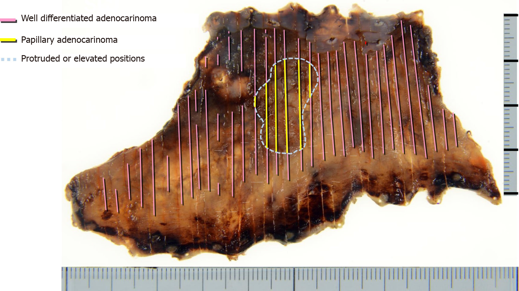
Figure 5 An endoscopic submucosal dissection en-bloc resected specimen and histological tumor distribution.
The superficial carcinoma consisted of papillary and well-differentiated tubular adenocarcinoma in the protruded and elevated positions and extensive flat portions, respectively. The adenocarcinoma lesion extended the whole circumference of the long-segment Barrett’s esophagus and was confined to the superficial muscularis mucosae.
- Citation: Abe K, Goda K, Kanamori A, Suzuki T, Yamamiya A, Takimoto Y, Arisaka T, Hoshi K, Sugaya T, Majima Y, Tominaga K, Iijima M, Hirooka S, Yamagishi H, Irisawa A. Whole circumferential endoscopic submucosal dissection of superficial adenocarcinoma in long-segment Barrett's esophagus: A case report. World J Gastrointest Surg 2021; 13(10): 1285-1292
- URL: https://www.wjgnet.com/1948-9366/full/v13/i10/1285.htm
- DOI: https://dx.doi.org/10.4240/wjgs.v13.i10.1285


