Copyright
©The Author(s) 2021.
World J Gastrointest Surg. Oct 27, 2021; 13(10): 1166-1179
Published online Oct 27, 2021. doi: 10.4240/wjgs.v13.i10.1166
Published online Oct 27, 2021. doi: 10.4240/wjgs.v13.i10.1166
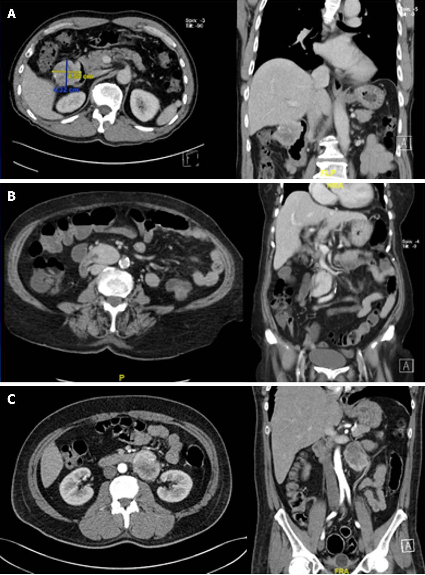
Figure 2 Computed tomography scan images of duodenal gastrointestinal stromal tumor in axial and coronal view.
A: A 4.58 cm × 4.32 cm heterogeneous enhancing mass located at the anti-mesenteric border of the second part of the duodenum; B: A 3.4 cm × 2.9 cm homogeneous enhancing lobulated soft tissue involving the third part of the duodenum showing both intra- and extraluminal component; C: An enhancing mixed density partly necrotic mass measuring 4.7 cm × 6.6 cm arising from the fourth part of the duodenum with a large exophytic component posteriorly.
- Citation: Lim KT. Current surgical management of duodenal gastrointestinal stromal tumors. World J Gastrointest Surg 2021; 13(10): 1166-1179
- URL: https://www.wjgnet.com/1948-9366/full/v13/i10/1166.htm
- DOI: https://dx.doi.org/10.4240/wjgs.v13.i10.1166


