Copyright
©The Author(s) 2020.
World J Gastrointest Surg. Sep 27, 2020; 12(9): 397-406
Published online Sep 27, 2020. doi: 10.4240/wjgs.v12.i9.397
Published online Sep 27, 2020. doi: 10.4240/wjgs.v12.i9.397
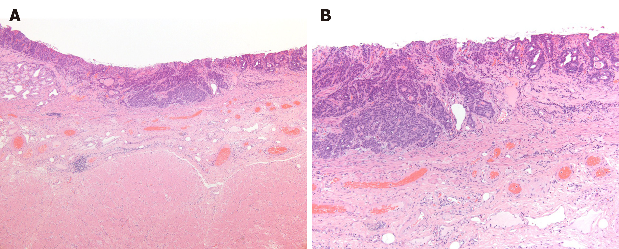
Figure 3 Pathological findings.
Pathological examination of the resected material revealed a moderately to poorly differentiated adenocarcinoma with submucosal invasion and ulcerative scars, but without lymphovascular invasion. A: Hematoxylin and eosin staining results, × 4; B: Hematoxylin and eosin staining results, × 10.
- Citation: Yura M, Koyanagi K, Adachi K, Hara A, Hayashi K, Tajima Y, Kaneko Y, Fujisaki H, Hirata A, Takano K, Hongo K, Yo K, Yoneyama K, Dehari R, Nakagawa M. Distal gastric tube resection with vascular preservation for gastric tube cancer: A case report and review of literature. World J Gastrointest Surg 2020; 12(9): 397-406
- URL: https://www.wjgnet.com/1948-9366/full/v12/i9/397.htm
- DOI: https://dx.doi.org/10.4240/wjgs.v12.i9.397


