Copyright
©The Author(s) 2019.
World J Gastrointest Surg. Jun 27, 2019; 11(6): 296-302
Published online Jun 27, 2019. doi: 10.4240/wjgs.v11.i6.296
Published online Jun 27, 2019. doi: 10.4240/wjgs.v11.i6.296
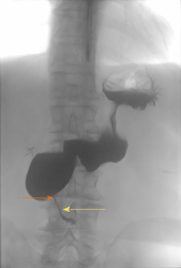
Figure 2 Upper gastrointestinal series showing the obstruction of duodenum.
The image shows the site of obstruction with retrograde dilation of duodenum (orange arrow). A very small portion of contrast agent passed through the obstruction (yellow arrow).
- Citation: Mikulić D, Bubalo T, Mrzljak A, Škrtić A, Jadrijević S, Kanižaj TF, Kocman B. Role of total pancreatectomy in the treatment of paraduodenal pancreatitis: A case report. World J Gastrointest Surg 2019; 11(6): 296-302
- URL: https://www.wjgnet.com/1948-9366/full/v11/i6/296.htm
- DOI: https://dx.doi.org/10.4240/wjgs.v11.i6.296


