Copyright
©The Author(s) 2019.
World J Gastrointest Surg. Feb 27, 2019; 11(2): 62-84
Published online Feb 27, 2019. doi: 10.4240/wjgs.v11.i2.62
Published online Feb 27, 2019. doi: 10.4240/wjgs.v11.i2.62
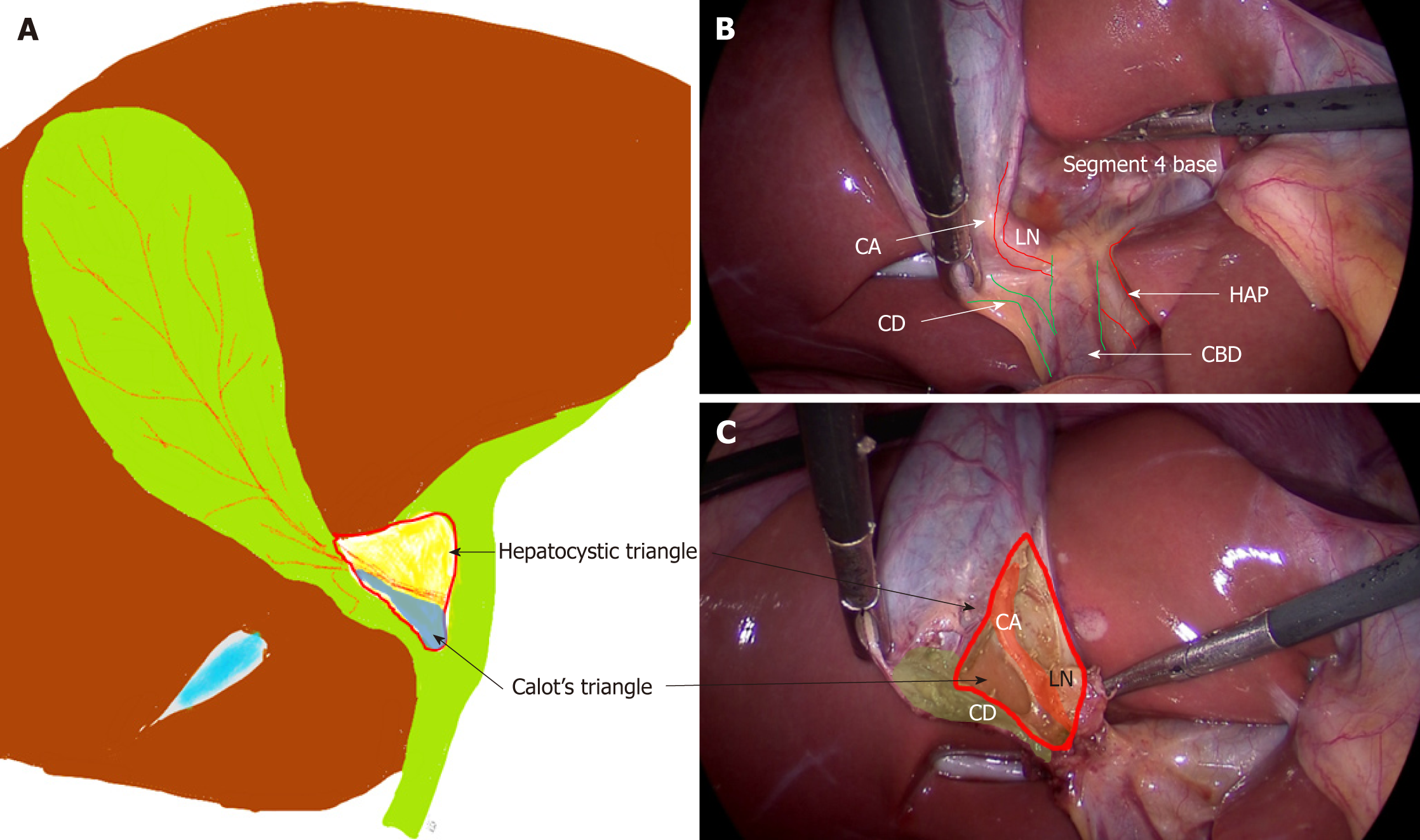
Figure 1 Anatomy of hepatocystic and Calot’s triangles.
A: Hepatocystic triangle (red outline); Calot’s triangle (in blue); Relevant anatomical structures; B: Before dissection; C: After dissection. CD: Cystic duct; CA: Cystic artery; LN: Lymph node; HAP: Hepatic artery proper; CBD: Common bile duct.
- Citation: Gupta V, Jain G. Safe laparoscopic cholecystectomy: Adoption of universal culture of safety in cholecystectomy. World J Gastrointest Surg 2019; 11(2): 62-84
- URL: https://www.wjgnet.com/1948-9366/full/v11/i2/62.htm
- DOI: https://dx.doi.org/10.4240/wjgs.v11.i2.62


