Copyright
©The Author(s) 2019.
World J Gastrointest Surg. Nov 27, 2019; 11(11): 414-421
Published online Nov 27, 2019. doi: 10.4240/wjgs.v11.i11.414
Published online Nov 27, 2019. doi: 10.4240/wjgs.v11.i11.414
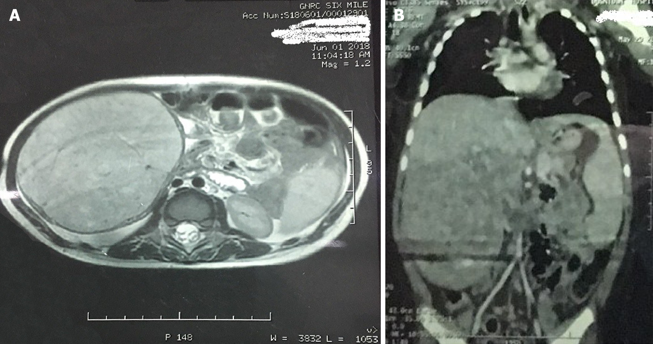
Figure 1 Magnetic resonance images.
A: Magnetic resonance imaging (cut section) shows the large mass arising from the inferior liver margin; B: Magnetic resonance imaging (coronal view) shows the large mass arising from the inferior liver margin.
- Citation: Roy AK, Das NN. Pediatric intrahepatic bile duct adenoma - rare liver tumor: A case report. World J Gastrointest Surg 2019; 11(11): 414-421
- URL: https://www.wjgnet.com/1948-9366/full/v11/i11/414.htm
- DOI: https://dx.doi.org/10.4240/wjgs.v11.i11.414


