Copyright
©The Author(s) 2015.
World J Gastrointest Surg. Dec 27, 2015; 7(12): 403-407
Published online Dec 27, 2015. doi: 10.4240/wjgs.v7.i12.403
Published online Dec 27, 2015. doi: 10.4240/wjgs.v7.i12.403
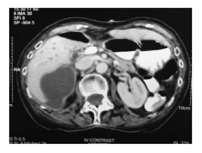
Figure 1 Axial section showing peripherally enhancing fluid density area seen along the segment 5 and 6 of the liver.
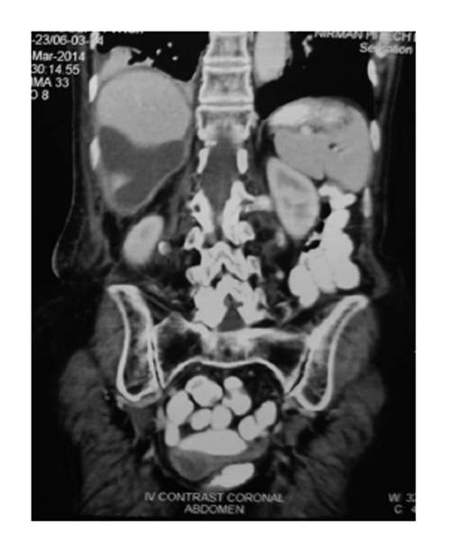
Figure 2 Coronal section showing peripherally enhancing fluid density area seen along the segment 6 of the liver.
The gall bladder is not seen in the gall bladder fossa.
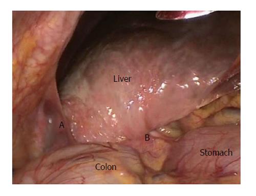
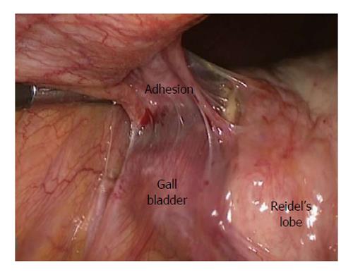
Figure 3 Laparoscopic view showing distorted anatomy.
A: Adhesions between the gall bladder and the lateral abdominal wall; B: Pulled in peritoneal fold from the pylorus of the stomach and the hepatic flexure.
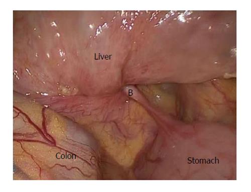
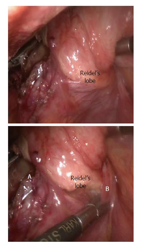
Figure 4 Zoomed in view of Calot’s region.
B: Pulled in peritoneal fold from the pylorus of the stomach and the hepatic flexure.
Figure 5 Adhesions between the gall bladder and the lateral abdominal wall.
Figure 6 Rotated Riedel’s lobe.
A: Gangrenous fundus of the gall bladder; B: Pulled in peritoneal fold from the pylorus of the stomach and the hepatic flexure.
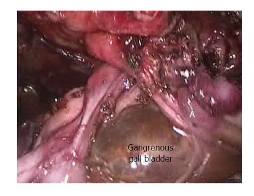
Figure 7 Gangrenous gall bladder after adhesiolysis.
A: Adhesive band between the neck of the gall bladder and the Riedel’s lobe.
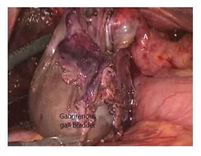
Figure 8 Gall bladder found gangrenous till its neck.
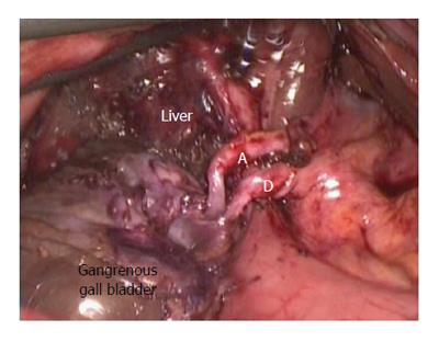
Figure 9 After derotation, the gall bladder in its normal position with the long and twisted cystic artery (A) and cystic duct (D).
- Citation: Sunder YK, Akhilesh SP, Raman G, Deborshi S, Shantilal MH. Laparoscopic management of a two staged gall bladder torsion. World J Gastrointest Surg 2015; 7(12): 403-407
- URL: https://www.wjgnet.com/1948-9366/full/v7/i12/403.htm
- DOI: https://dx.doi.org/10.4240/wjgs.v7.i12.403


