Copyright
©The Author(s) 2015.
World J Diabetes. May 15, 2015; 6(4): 662-672
Published online May 15, 2015. doi: 10.4239/wjd.v6.i4.662
Published online May 15, 2015. doi: 10.4239/wjd.v6.i4.662
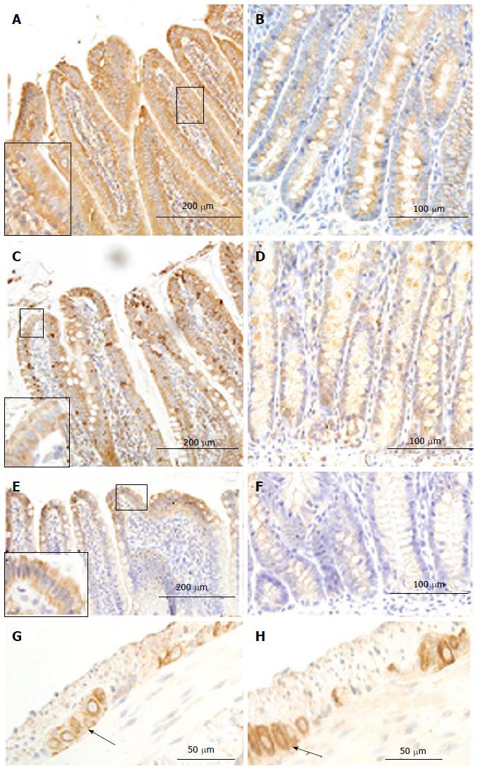
Figure 6 Receptor of advanced glycation end products immune-staining in villi (A, C, E) and crypt (B, D, F) of duodenum (A, B), jejunum (C, D) and ileum(E, F) as well as in ileum ganglia (arrowhead; G: Normal group; H: GK group).
As shown in the magnification area (big frame vs small frame), the RAGE homogeneously distributed in the epithelia cells. The intensity of immune-staining in ganglia was stronger in the diabetic group (H) than in the normal group (G) (arrowhead). RAGE: Receptor of advanced glycation end products; GK: Inherited type 2 diabetic Goto-Kakizak rats.
- Citation: Chen PM, Gregersen H, Zhao JB. Advanced glycation end-product expression is upregulated in the gastrointestinal tract of type 2 diabetic rats. World J Diabetes 2015; 6(4): 662-672
- URL: https://www.wjgnet.com/1948-9358/full/v6/i4/662.htm
- DOI: https://dx.doi.org/10.4239/wjd.v6.i4.662


