
















©2014 Baishideng Publishing Group Co.
World J Diabetes. Feb 15, 2014; 5(1): 59-68
Published online Feb 15, 2014. doi: 10.4239/wjd.v5.i1.59
Published online Feb 15, 2014. doi: 10.4239/wjd.v5.i1.59
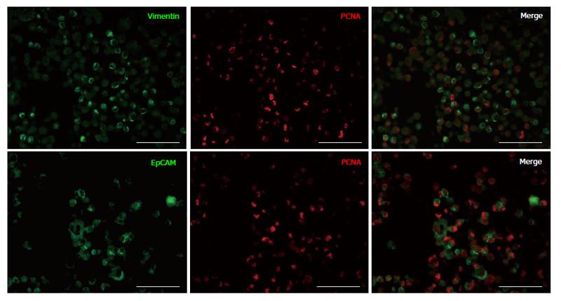
Figure 1 Double immunofluorescence staining of islet depleted pancreatic tissue expanded cells after 14 d in culture.
Cells are positive for vimentin (green), EpCAM (green) and PCNA (red). Both vimentin and EpCAM positive cells are positive for PCNA staining (merged) and proliferating. Scale bars are 50 μm. EpCAM: Epithelial cell adhesion molecule; PCNA: Proliferating cell nuclear antigen.
- Citation: Seeberger KL, Anderson SJ, Ellis CE, Yeung TY, Korbutt GS. Identification and differentiation of PDX1 β-cell progenitors within the human pancreatic epithelium. World J Diabetes 2014; 5(1): 59-68
- URL: https://www.wjgnet.com/1948-9358/full/v5/i1/59.htm
- DOI: https://dx.doi.org/10.4239/wjd.v5.i1.59