
















Copyright: ©Author(s) 2026.
World J Diabetes. Sep 15, 2026; 17(9): 122779
Published online Sep 15, 2026. doi: 10.4239/wjd.122779
Published online Sep 15, 2026. doi: 10.4239/wjd.122779
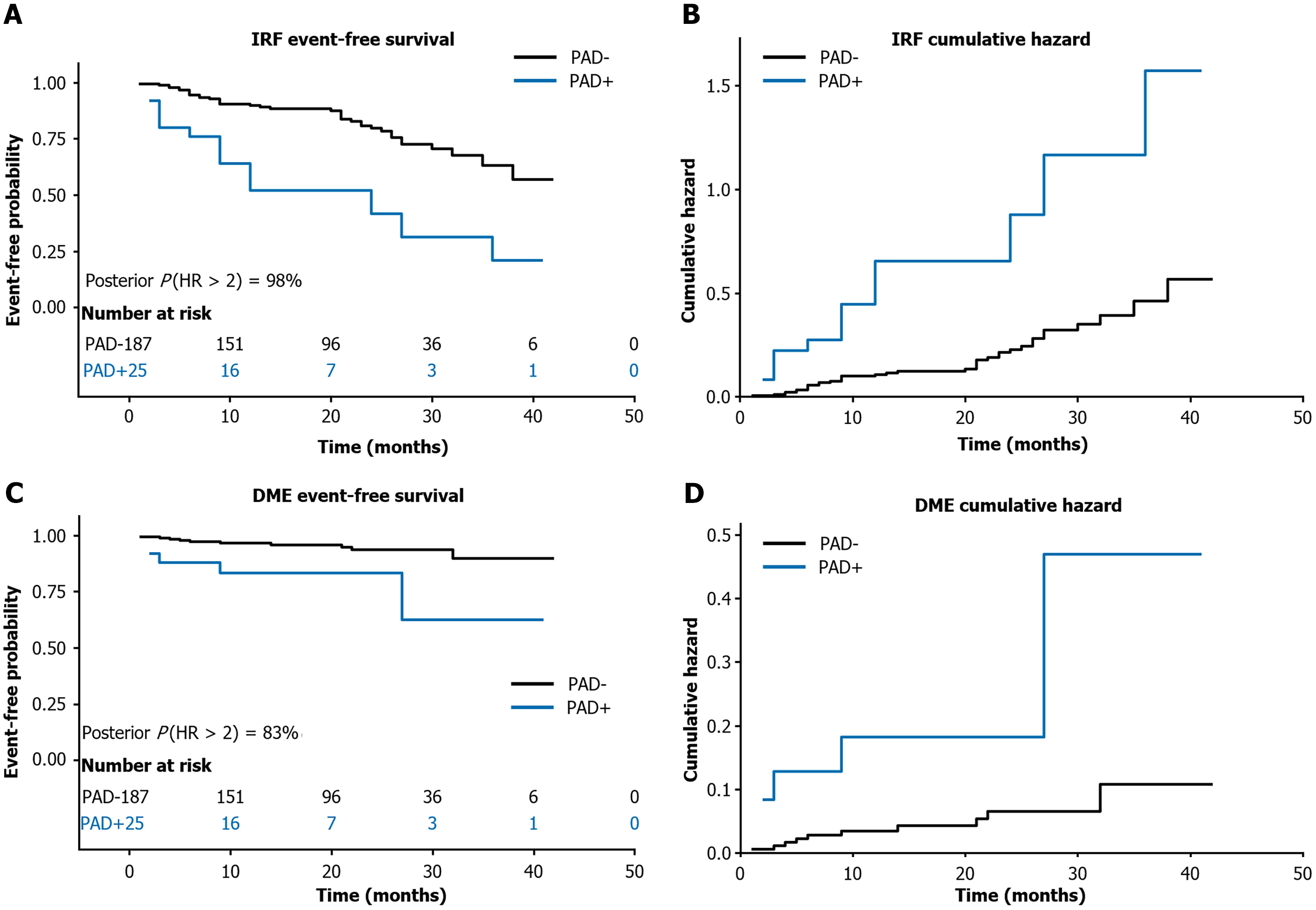
Figure 2 Kaplan-Meier survival curves by peripheral arterial disease status.
A: Intra-retinal fluid event-free survival; B: Intra-retinal fluid cumulative hazard; C: Diabetic macular edema event-free survival; D: Diabetic macular edema cumulative hazard. Black = peripheral arterial disease- (n = 187); blue = peripheral arterial disease + (n = 25). IRF: Intra-retinal fluid; DME: Diabetic macular edema; HR: Hazard ratio; PAD: Peripheral arterial disease.
- Citation: Yang JM, Kwak J, Min SH, Choi SU, Park J, Yon DK, Lee JY, Kim JG, Yoon YH, Kim YJ. Peripheral arterial disease is associated with retinal fluid and microvascular alterations in type 2 diabetes. World J Diabetes 2026; 17(9): 122779
- URL: https://www.wjgnet.com/1948-9358/full/v17/i9/122779.htm
- DOI: https://dx.doi.org/10.4239/wjd.122779