
















Copyright: ©Author(s) 2026.
World J Diabetes. Sep 15, 2026; 17(9): 120935
Published online Sep 15, 2026. doi: 10.4239/wjd.120935
Published online Sep 15, 2026. doi: 10.4239/wjd.120935
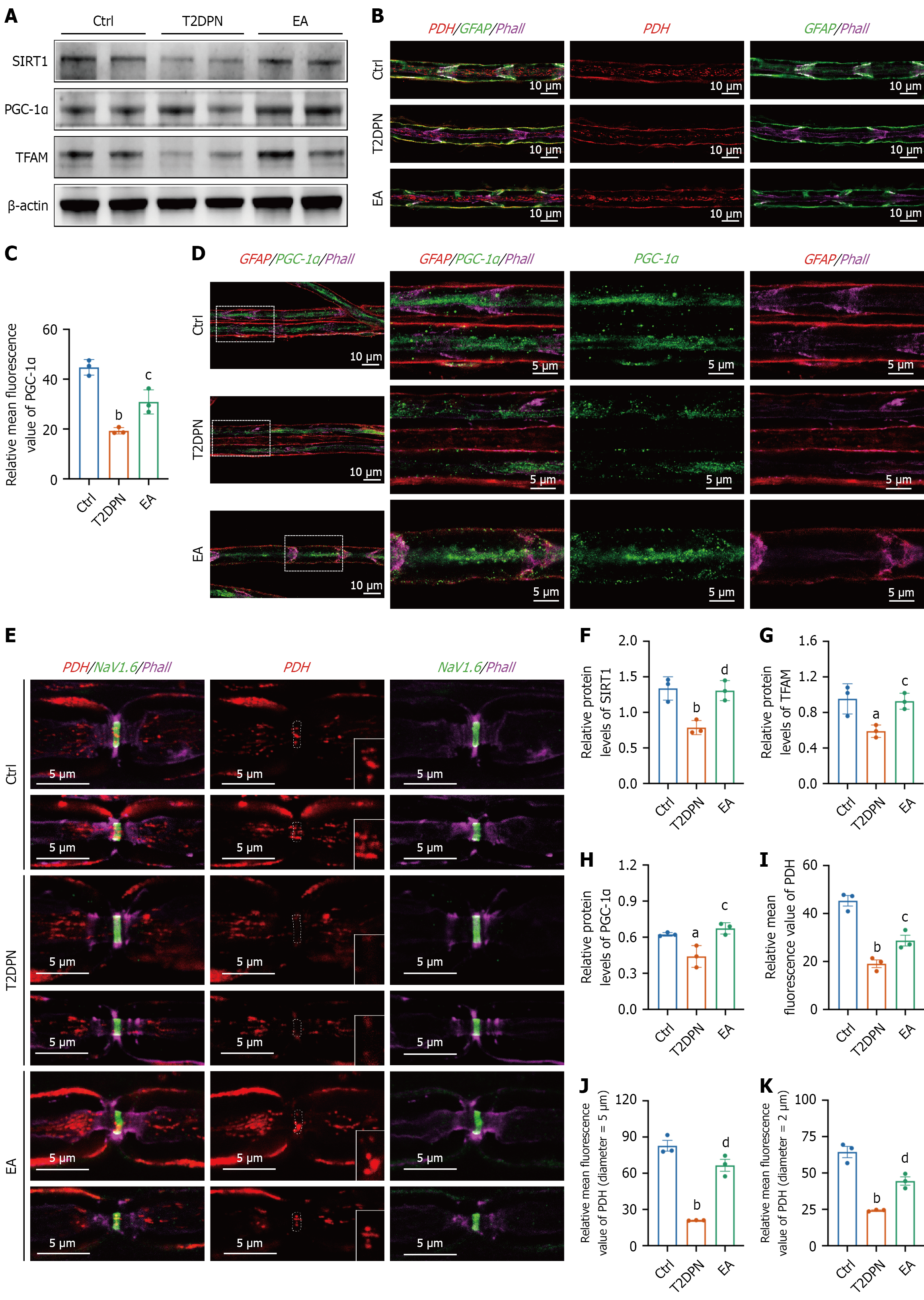
Figure 4 Electroacupuncture treatment upregulated the silent information regulator 1/peroxisome proliferator-activated receptor-γ coactivator-1α pathway in the sciatic nerve of type 2 diabetic peripheral neuropathy rats.
A: Western blot analysis of silent information regulator 1 (SIRT1), peroxisome proliferator-activated receptor-γ coactivator-1α (PGC-1α), and mitochondrial transcription factor A (TFAM) in sciatic nerve tissue; B: Representative immunofluorescence images of pyruvate dehydrogenase (PDH)-stained mitochondria (red) in sciatic nerve fibers; C: Quantification of the mean fluorescence intensity of PGC-1α staining in sciatic nerve fibers; D: Representative immunofluorescence images of PGC-1α staining (green) in sciatic nerve fibers; E: Representative immunofluorescence images of PDH-stained mitochondria (red) at nodes of Ranvier marked by voltage-gated sodium channel 1.6 (NaV1.6) (green) in sciatic nerve fibers with axonal diameters of 5 μm and 2 μm. The white dotted frames indicate the positions of nodes of Ranvier, and the 2 × magnified images show mitochondria at the nodes of Ranvier; F-H: Quantitative analysis of SIRT1 (F), TFAM (G), and PGC-1α (H) protein expression in sciatic nerve tissue; I: Quantification of the mean fluorescence intensity of PDH-stained mitochondria in sciatic nerve fibers; J and K: Quantification of the relative mean fluorescence intensity of mitochondria at nodes of Ranvier in axons with diameters of 5 μm (J) and 2 μm (K). Glial fibrillary acidic protein was stained for glial cells (green in B and red in D), phalloidin was stained for axons (magenta in B, D, and E), and NaV1.6 was stained for nodes of Ranvier (green in E). Data are presented as mean ± SEM (n = 3). aP < 0.05 type 2 diabetic peripheral neuropathy group vs control group. bP < 0.01 type 2 diabetic peripheral neuropathy group vs control group. cP < 0.05 electroacupuncture group vs type 2 diabetic peripheral neuropathy group. dP < 0.01 electroacupuncture group vs type 2 diabetic peripheral neuropathy group. T2DPN: Type 2 diabetic peripheral neuropathy; EA: Electroacupuncture; Ctrl: Control; SIRT1: Silent information regulator 1; PGC-1α: Peroxisome proliferator-activated receptor-γ coactivator-1α; TFAM: Mitochondrial transcription factor A; Phall: Phalloidin; PDH: Pyruvate dehydrogenase; GFAP: Glial fibrillary acidic protein; NaV1.6: Voltage-gated sodium channel 1.6.
- Citation: Wang X, Yuan CX, Liu Y, Li WF, Lu MJ, Xu TC, Yu Z, Xu B. Electroacupuncture alleviates diabetic peripheral neuropathy: Regulates mitochondrial biogenesis and redistribution in peripheral nerve fibers. World J Diabetes 2026; 17(9): 120935
- URL: https://www.wjgnet.com/1948-9358/full/v17/i9/120935.htm
- DOI: https://dx.doi.org/10.4239/wjd.120935