
















Copyright: ©Author(s) 2026.
World J Diabetes. Sep 15, 2026; 17(9): 120935
Published online Sep 15, 2026. doi: 10.4239/wjd.120935
Published online Sep 15, 2026. doi: 10.4239/wjd.120935
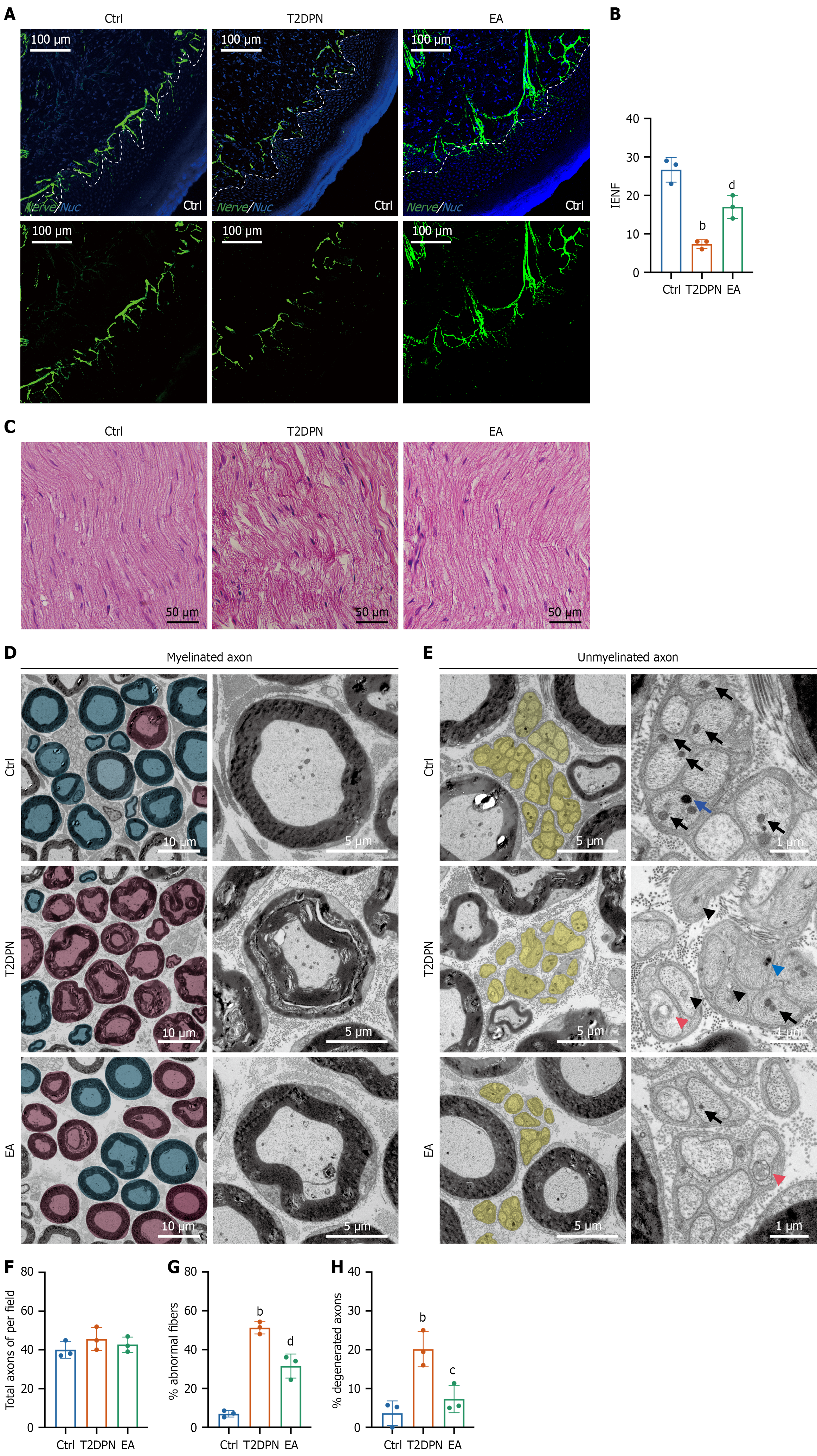
Figure 3 Electroacupuncture treatment increases intraepidermal nerve fiber density and ameliorates degeneration of sciatic nerve fibers in type 2 diabetic peripheral neuropathy rats.
A: Representative images of protein gene product 9.5-positive intraepidermal nerve fibers (green) and 4’,6-diamidino-2-phenylindole staining (blue) in hind paw skin; B: Quantification of intraepidermal nerve fiber density in different groups. Data are presented as mean ± SEM (n = 3); C: Longitudinal hematoxylin-eosin-stained sections of the sciatic nerve in different groups; D: Representative transmission electron microscopy images of myelinated axons with pseudo-coloring of intact axons (blue mask) and degenerated axons (pink mask) (left), and magnified individual axons (right) in sciatic nerve sections from different groups; E: Representative images of unmyelinated axons (yellow mask) (left) and magnified Remak bundles (right) in sciatic nerve sections from different groups. Normal and damaged mitochondria are indicated by black arrows and arrowheads, respectively. Normal and increased lysosomes are indicated by blue arrows and arrowheads, respectively. Autophagosomes are indicated by magenta arrowheads; F: Related morphological analyses of axons per field; G: Percentage of abnormal fibers; H: Percentage of degenerated axons in different groups based on myelinated axons. Data are presented as mean ± SEM (n = 3). bP < 0.01 type 2 diabetic peripheral neuropathy group vs control group. cP < 0.05 electroacupuncture group vs type 2 diabetic peripheral neuropathy group. dP < 0.01 electroacupuncture group vs type 2 diabetic peripheral neuropathy group. T2DPN: Type 2 diabetic peripheral neuropathy; EA: Electroacupuncture; Ctrl: Control; IENF: Intraepidermal nerve fibers.
- Citation: Wang X, Yuan CX, Liu Y, Li WF, Lu MJ, Xu TC, Yu Z, Xu B. Electroacupuncture alleviates diabetic peripheral neuropathy: Regulates mitochondrial biogenesis and redistribution in peripheral nerve fibers. World J Diabetes 2026; 17(9): 120935
- URL: https://www.wjgnet.com/1948-9358/full/v17/i9/120935.htm
- DOI: https://dx.doi.org/10.4239/wjd.120935