
















Copyright: ©Author(s) 2026.
World J Diabetes. Jul 15, 2026; 17(7): 120448
Published online Jul 15, 2026. doi: 10.4239/wjd.120448
Published online Jul 15, 2026. doi: 10.4239/wjd.120448
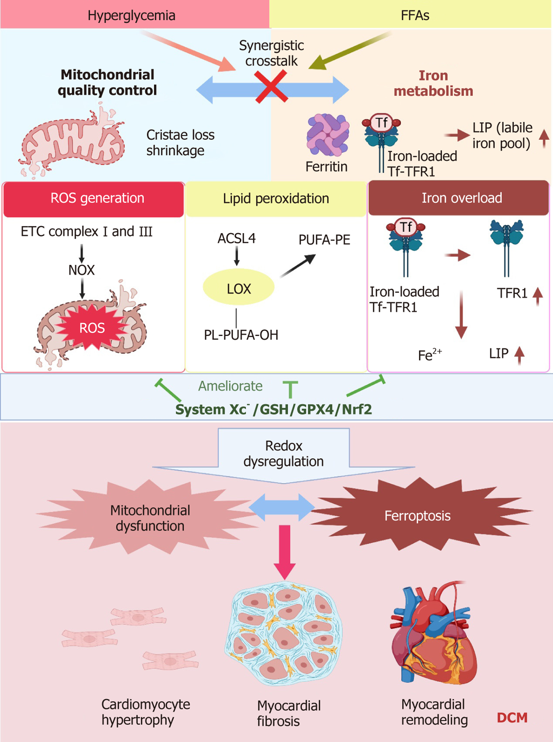
Figure 3 Crosstalk between mitochondrial dysfunction and ferroptosis in the pathogenesis of diabetic cardiomyopathy.
This schematic depicts the interplay between mitochondrial quality control and iron metabolism in diabetic cardiomyopathy (DCM). Hyperglycemia and elevated free fatty acids impair mitochondrial cristae integrity and enhance reactive oxygen species generation, whereas excessive iron uptake and dysregulated storage cause iron overload and expansion of the labile iron pool. These alterations promote lipid peroxidation via ACSL4/LOX pathways and disrupt redox homeostasis, thereby linking mitochondrial dysfunction with ferroptosis. Antioxidant defense mechanisms - including System Xc-, glutathione, GPX4, and Nrf2 - counteract ferroptotic stress. The synergistic interaction between mitochondrial impairment and ferroptosis drives cardiomyocyte hypertrophy, myocardial fibrosis, and adverse cardiac remodeling, ultimately accelerating the progression of DCM. FFAs: Free fatty acids; ETC: Electron transport chain; NOX: NADPH oxidase; DCM: Diabetic cardiomyopathy; ROS: Reactive oxygen species; GSH: Glutathione.
- Citation: Tang YT, Wu Q, Chen YP, Xia L, Yang MH, Wei MY, Pang Q, Yang YN, Liu JB, Liu JL, Ni Q, Gong YB. Regulation of ferroptosis and mitochondrial homeostasis disruption in diabetic cardiomyopathy: Therapeutic potential of traditional Chinese medicine. World J Diabetes 2026; 17(7): 120448
- URL: https://www.wjgnet.com/1948-9358/full/v17/i7/120448.htm
- DOI: https://dx.doi.org/10.4239/wjd.120448