
















Copyright: ©Author(s) 2026.
World J Diabetes. Jun 15, 2026; 17(6): 121606
Published online Jun 15, 2026. doi: 10.4239/wjd.121606
Published online Jun 15, 2026. doi: 10.4239/wjd.121606
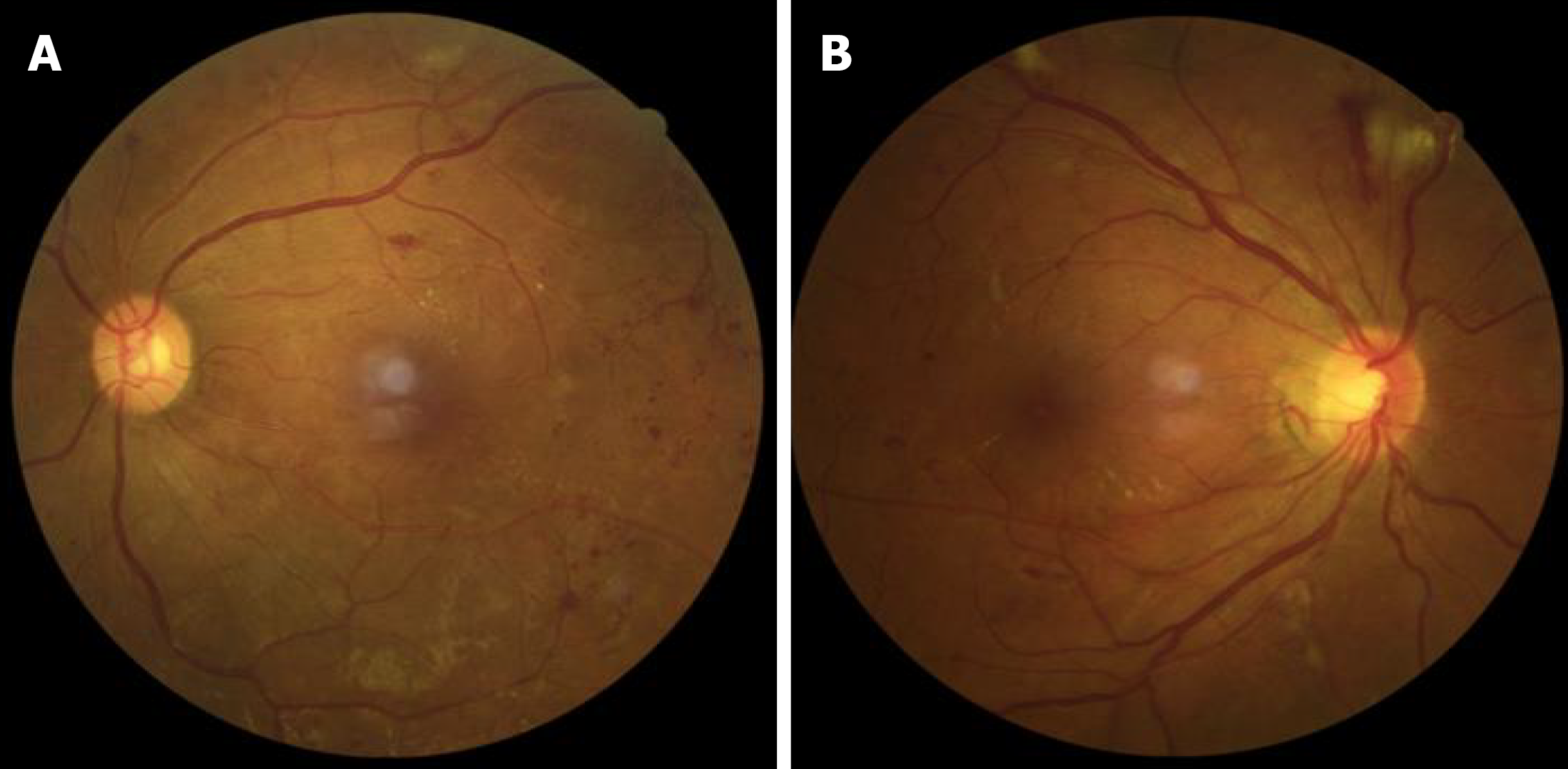
Figure 3 Color fundus photography of the patient.
A: Left eye; B: Right eye. Both images demonstrate characteristic features of proliferative diabetic retinopathy, including retinal hemorrhages, exudates, and neovascularization.
- Citation: Li GZ, Liu JY, Hao HY, Zhou H. Clinical mismatches leading to the misdiagnosis of multiple myeloma as diabetic nephropathy: A case report. World J Diabetes 2026; 17(6): 121606
- URL: https://www.wjgnet.com/1948-9358/full/v17/i6/121606.htm
- DOI: https://dx.doi.org/10.4239/wjd.121606